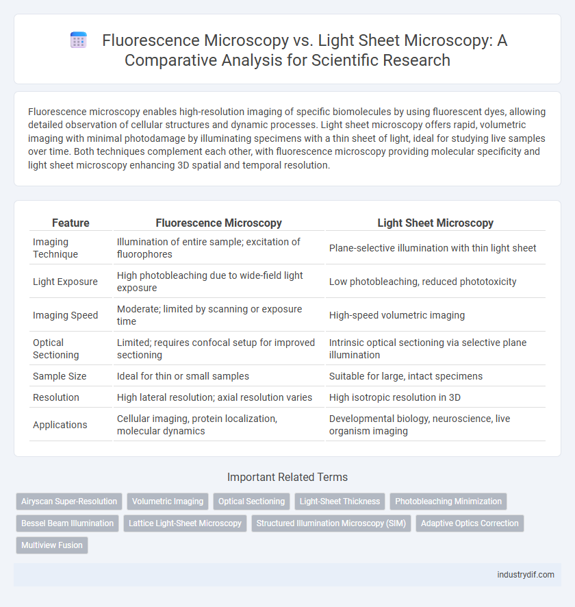
Fluorescence microscopy enables high-resolution imaging of specific biomolecules by using fluorescent dyes, allowing detailed observation of cellular structures and dynamic processes. Light sheet microscopy offers rapid, volumetric imaging with minimal photodamage by illuminating specimens with a thin sheet of light, ideal for studying live samples over time. Both techniques complement each other, with fluorescence microscopy providing molecular specificity and light sheet microscopy enhancing 3D spatial and temporal resolution.
Table of Comparison
| Feature | Fluorescence Microscopy | Light Sheet Microscopy |
|---|---|---|
| Imaging Technique | Illumination of entire sample; excitation of fluorophores | Plane-selective illumination with thin light sheet |
| Light Exposure | High photobleaching due to wide-field light exposure | Low photobleaching, reduced phototoxicity |
| Imaging Speed | Moderate; limited by scanning or exposure time | High-speed volumetric imaging |
| Optical Sectioning | Limited; requires confocal setup for improved sectioning | Intrinsic optical sectioning via selective plane illumination |
| Sample Size | Ideal for thin or small samples | Suitable for large, intact specimens |
| Resolution | High lateral resolution; axial resolution varies | High isotropic resolution in 3D |
| Applications | Cellular imaging, protein localization, molecular dynamics | Developmental biology, neuroscience, live organism imaging |
Introduction to Fluorescence Microscopy
Fluorescence microscopy utilizes fluorescent dyes or proteins to label specific cellular components, enabling high-contrast imaging of biological samples. It relies on excitation light to induce fluorescence emission, which is collected to produce detailed images of structures within cells and tissues. This technique offers spatial resolution and molecular specificity critical for studying dynamic processes in live specimens.
Fundamentals of Light Sheet Microscopy
Light Sheet Microscopy (LSM) utilizes a thin sheet of laser light to selectively illuminate a single plane within a specimen, minimizing phototoxicity and photobleaching compared to traditional Fluorescence Microscopy. This orthogonal illumination approach enables high-speed, volumetric imaging with improved optical sectioning and reduced background fluorescence. The fundamental advantage of LSM lies in decoupling the illumination and detection pathways, enhancing spatial resolution and allowing for long-term live-cell imaging with minimal damage.
Principles of Image Formation
Fluorescence microscopy relies on the excitation of fluorophores within a specimen using specific wavelengths of light, causing them to emit light at longer wavelengths which are then detected to form an image. Light sheet microscopy forms images by illuminating a thin plane of the specimen with a sheet of light, allowing optical sectioning and reducing photobleaching and phototoxicity. This method enables high-speed, high-contrast imaging of thick biological samples by capturing fluorescence signals from the illuminated plane orthogonally with a separate detection objective.
Spatial Resolution Comparison
Fluorescence microscopy typically achieves a spatial resolution of approximately 200 nm laterally and 500 nm axially, constrained by the diffraction limit of light. Light sheet microscopy offers improved axial resolution and reduced photobleaching by illuminating a thin plane within the specimen, allowing optical sectioning with high contrast. Comparative studies reveal that light sheet microscopy enhances volumetric imaging of live samples while maintaining similar lateral resolution to conventional fluorescence techniques.
Sample Preparation Techniques
Fluorescence microscopy typically requires extensive sample preparation including fixation, permeabilization, and fluorescent labeling to enhance signal specificity and reduce background noise. Light sheet microscopy benefits from gentle sample handling with minimal photodamage, often utilizing cleared or optically transparent specimens to enable deep tissue imaging. Both techniques rely on optimized mounting strategies and refractive index matching to improve image quality and resolution.
Photobleaching and Phototoxicity
Fluorescence microscopy often induces significant photobleaching and phototoxicity due to the intense, focused illumination required for excitation, which can damage live samples and limit imaging duration. In contrast, light sheet microscopy reduces photobleaching and phototoxicity by illuminating only a thin plane of the specimen, minimizing light exposure and preserving sample viability during extended imaging sessions. This selective plane illumination is particularly advantageous for long-term live-cell imaging and high-resolution volumetric analysis in biological research.
Imaging Speed and Throughput
Fluorescence microscopy offers high spatial resolution but is often limited by slower imaging speed and lower throughput due to point-by-point scanning methods. Light sheet microscopy significantly enhances imaging speed and throughput by illuminating a thin plane of the specimen, enabling rapid acquisition of large volumetric datasets with minimal photodamage. This makes light sheet microscopy particularly advantageous for live-cell imaging and high-content screening applications requiring fast, high-throughput data collection.
Applications in Biological Research
Fluorescence microscopy enables high-resolution imaging of specific biomolecules within cells by exploiting fluorescent tags, making it indispensable for studying cellular structures, protein localization, and dynamic processes in fixed and live samples. Light sheet microscopy offers fast volumetric imaging with minimal phototoxicity, allowing researchers to visualize large, living organisms and developmental processes in real time. Combining these techniques enhances biological research by providing complementary insights into molecular interactions and three-dimensional tissue architecture.
Limitations and Challenges
Fluorescence microscopy faces limitations such as photobleaching, limited penetration depth, and phototoxicity, which restrict prolonged live-cell imaging and imaging of thick specimens. Light sheet microscopy overcomes some depth and photodamage issues by illuminating specimens with a thin sheet of light, but challenges include complex setup, restricted sample size, and optical aberrations causing reduced resolution at deeper tissue layers. Both techniques demand careful optimization of fluorophore selection and imaging parameters to balance resolution, speed, and sample viability.
Future Directions in Microscopy Technologies
Fluorescence microscopy continues to evolve with innovations in super-resolution techniques and adaptive optics to enhance spatial resolution and live-cell imaging capabilities. Light sheet microscopy is pushing boundaries by integrating faster volumetric imaging and multi-scale light-sheet designs to minimize phototoxicity while capturing dynamic biological processes in real time. Emerging trends in microscopy technologies emphasize combining fluorescence sensitivity with light sheet's gentle illumination to enable high-throughput, deep-tissue, and long-term imaging in complex biological systems.
Related Important Terms
Airyscan Super-Resolution
Airyscan super-resolution microscopy enhances spatial resolution and signal-to-noise ratio beyond conventional fluorescence microscopy by utilizing a detector array for improved photon collection, enabling detailed cellular imaging at sub-diffraction limits. Compared to light sheet microscopy, Airyscan provides higher lateral and axial resolution for thin optical sections but illuminates the entire sample volume, whereas light sheet microscopy minimizes photodamage by selective plane illumination ideal for large, living specimens over extended time-lapse imaging.
Volumetric Imaging
Fluorescence microscopy enables high-resolution imaging of specific biomolecules but is often limited by photobleaching and slower volumetric acquisition. Light sheet microscopy offers rapid volumetric imaging with reduced phototoxicity, allowing long-term observation of dynamic biological processes in three dimensions.
Optical Sectioning
Fluorescence microscopy offers optical sectioning by exciting fluorophores in a single focal plane, but suffers from out-of-focus light that degrades image contrast and resolution. Light sheet microscopy enhances optical sectioning by illuminating the sample with a thin sheet of light orthogonal to the detection axis, reducing photobleaching and improving signal-to-noise ratio in three-dimensional imaging.
Light-Sheet Thickness
Light sheet microscopy offers significantly reduced light-sheet thickness, typically ranging from 1 to 10 micrometers, enabling high axial resolution and minimizing phototoxicity compared to conventional fluorescence microscopy, which generally has thicker light sheets due to point illumination. This ultrathin illumination in light sheet microscopy enhances optical sectioning capability and preserves sample integrity during prolonged imaging sessions.
Photobleaching Minimization
Light sheet microscopy significantly reduces photobleaching by illuminating only a thin plane of the specimen, unlike fluorescence microscopy which exposes the entire sample to excitation light, causing widespread photodamage. This targeted illumination enhances imaging longevity and preserves sample integrity during prolonged observation of dynamic biological processes.
Bessel Beam Illumination
Bessel beam illumination in light sheet microscopy provides enhanced axial resolution and reduced photobleaching compared to conventional fluorescence microscopy by generating a non-diffracting, self-reconstructing light sheet that enables deeper tissue imaging with minimal phototoxicity. This technique improves contrast and imaging speed, making it particularly advantageous for live-cell and long-term dynamic studies in complex biological specimens.
Lattice Light-Sheet Microscopy
Lattice Light-Sheet Microscopy (LLSM) offers high spatiotemporal resolution and reduced phototoxicity compared to traditional fluorescence microscopy by illuminating samples with a thin sheet of patterned light. This technique enables rapid volumetric imaging of live cells and organisms with minimal photobleaching, enhancing dynamic biological process visualization at subcellular levels.
Structured Illumination Microscopy (SIM)
Structured Illumination Microscopy (SIM) enhances fluorescence microscopy by surpassing diffraction limits, enabling super-resolution imaging with doubled spatial resolution compared to conventional widefield techniques. Unlike light sheet microscopy, which illuminates samples plane-by-plane for optical sectioning, SIM employs patterned illumination and computational reconstruction to improve resolution and contrast in fluorescence imaging without requiring specialized sample mounting or geometric constraints.
Adaptive Optics Correction
Adaptive optics correction in fluorescence microscopy enhances image resolution by compensating for optical aberrations caused by sample heterogeneity, enabling improved visualization of subcellular structures in thick tissues. In light sheet microscopy, adaptive optics correction optimizes illumination and detection paths, significantly reducing scattering and photodamage while maintaining high-speed volumetric imaging performance.
Multiview Fusion
Fluorescence microscopy provides high-resolution imaging of fluorescently labeled samples but often suffers from photobleaching and limited penetration depth, whereas light sheet microscopy enables rapid, volumetric imaging with reduced phototoxicity by illuminating samples with a thin sheet of light. Multiview fusion in light sheet microscopy enhances spatial resolution and isotropy by computationally integrating multiple angled views, overcoming optical aberrations and sample scattering challenges inherent in single-view fluorescence microscopy.
Fluorescence Microscopy vs Light Sheet Microscopy Infographic

 industrydif.com