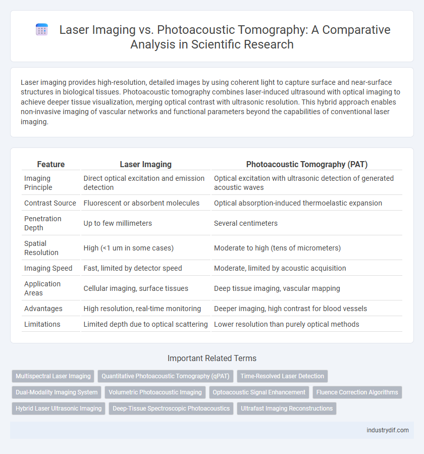
Laser imaging provides high-resolution, detailed images by using coherent light to capture surface and near-surface structures in biological tissues. Photoacoustic tomography combines laser-induced ultrasound with optical imaging to achieve deeper tissue visualization, merging optical contrast with ultrasonic resolution. This hybrid approach enables non-invasive imaging of vascular networks and functional parameters beyond the capabilities of conventional laser imaging.
Table of Comparison
| Feature | Laser Imaging | Photoacoustic Tomography (PAT) |
|---|---|---|
| Imaging Principle | Direct optical excitation and emission detection | Optical excitation with ultrasonic detection of generated acoustic waves |
| Contrast Source | Fluorescent or absorbent molecules | Optical absorption-induced thermoelastic expansion |
| Penetration Depth | Up to few millimeters | Several centimeters |
| Spatial Resolution | High (<1 um in some cases) | Moderate to high (tens of micrometers) |
| Imaging Speed | Fast, limited by detector speed | Moderate, limited by acoustic acquisition |
| Application Areas | Cellular imaging, surface tissues | Deep tissue imaging, vascular mapping |
| Advantages | High resolution, real-time monitoring | Deeper imaging, high contrast for blood vessels |
| Limitations | Limited depth due to optical scattering | Lower resolution than purely optical methods |
Fundamental Principles of Laser Imaging
Laser imaging relies on the interaction of coherent light with biological tissues to generate high-resolution images based on light absorption, scattering, and reflection properties. This method utilizes lasers to produce focused light beams that penetrate tissues, enabling visualization of structural and functional information at cellular or subcellular levels. Laser imaging systems often employ techniques like confocal microscopy or optical coherence tomography to enhance image contrast and spatial resolution by detecting backscattered or fluorescent signals.
Core Mechanisms in Photoacoustic Tomography
Photoacoustic tomography (PAT) combines optical absorption contrast and ultrasonic resolution by detecting ultrasound waves generated via thermoelastic expansion after pulsed laser irradiation. This technique relies on the photoacoustic effect where short laser pulses cause rapid localized heating, inducing pressure waves detectable by ultrasonic transducers. The core mechanism enables high-contrast imaging of biological tissues by mapping optical absorption with ultrasonic spatial resolution, surpassing limitations of pure laser imaging.
Comparative Image Resolution Capabilities
Laser Imaging provides high spatial resolution by utilizing coherent light to capture surface details with micron-level precision, ideal for superficial tissue visualization. Photoacoustic Tomography combines optical absorption contrast with ultrasonic detection, enabling deeper tissue imaging but with comparatively lower spatial resolution than laser imaging. The trade-off between penetration depth and resolution differentiates laser imaging's fine surface detail capabilities from photoacoustic tomography's volumetric and functional imaging potential.
Depth Penetration and Tissue Interaction
Laser imaging offers limited depth penetration typically around 1-2 mm in biological tissues due to strong light scattering, restricting its utility for deep tissue visualization. Photoacoustic tomography (PAT) combines optical absorption contrast with ultrasonic spatial resolution, enabling imaging depths up to several centimeters by converting absorbed optical energy into acoustic waves that experience less scattering. Tissue interaction in PAT involves excitation of endogenous chromophores like hemoglobin, providing functional and molecular information beyond the structural imaging capabilities of conventional laser imaging.
Sensitivity and Specificity in Biological Imaging
Laser imaging techniques demonstrate high sensitivity in detecting cellular structures by using coherent light to generate detailed images based on optical scattering. Photoacoustic tomography combines optical absorption contrast with ultrasonic detection, offering superior specificity in differentiating tissue types due to its sensitivity to hemoglobin concentration and oxygenation levels. The integration of laser imaging's high spatial resolution with photoacoustic tomography's molecular specificity enhances overall diagnostic accuracy in biological imaging applications.
Applications in Medical Diagnostics
Laser imaging provides high-resolution visualization of superficial tissues, making it ideal for dermatological assessments and vascular imaging. Photoacoustic tomography combines optical contrast with ultrasonic detection, enabling deep tissue imaging and functional analysis of oxygen saturation and hemoglobin concentration. This hybrid modality enhances early cancer detection, brain imaging, and cardiovascular diagnostics by offering both anatomical and molecular information.
Safety Profiles and Patient Considerations
Laser imaging employs non-ionizing radiation, minimizing risks of tissue damage and making it generally safe for repeated use in clinical settings. Photoacoustic tomography combines laser pulses with ultrasound detection, providing deeper tissue visualization while maintaining low thermal exposure and minimal phototoxicity. Patient considerations include sensitivity to light exposure, potential for mild discomfort, and suitability for individuals with implanted medical devices or photosensitive conditions.
Advancements in Instrumentation and Technology
Recent advancements in laser imaging and photoacoustic tomography have significantly enhanced resolution and imaging depth through the integration of ultra-fast pulsed lasers and highly sensitive ultrasound transducers. Innovations in laser sources, including tunable wavelength lasers and fiber lasers, have improved tissue contrast and specificity in photoacoustic imaging, enabling detailed visualization of molecular and functional information. The development of scalable and miniaturized instrument architectures facilitates real-time, non-invasive imaging in clinical and preclinical applications, broadening the scope and precision of biomedical diagnostics.
Integration with Multimodal Imaging Systems
Laser imaging offers high spatial resolution and real-time visualization, making it effective for structural analysis in multimodal imaging systems. Photoacoustic tomography (PAT) integrates optical absorption contrast with ultrasonic detection, enabling deeper tissue imaging and functional assessment alongside anatomical data. Combining laser imaging and PAT enhances diagnostic accuracy by providing complementary information on tissue morphology and molecular composition within integrated biomedical platforms.
Future Trends in Noninvasive Imaging Technologies
Laser imaging and photoacoustic tomography (PAT) are advancing rapidly as noninvasive imaging technologies with promising applications in biomedical diagnostics. Future trends emphasize enhanced spatial resolution, deeper tissue penetration, and multimodal integration combining optical and acoustic contrasts to improve functional and molecular imaging. Emerging developments in laser source miniaturization, real-time data processing, and AI-driven image reconstruction will further enable precise, noninvasive visualization of complex biological structures.
Related Important Terms
Multispectral Laser Imaging
Multispectral Laser Imaging leverages multiple laser wavelengths to capture detailed spectral information, enabling precise tissue characterization beyond standard Laser Imaging techniques. Photoacoustic Tomography combines laser-induced ultrasound signals with optical absorption contrast, providing deeper tissue visualization but with less spectral specificity compared to Multispectral Laser Imaging.
Quantitative Photoacoustic Tomography (qPAT)
Quantitative Photoacoustic Tomography (qPAT) integrates optical absorption and acoustic wave detection to provide high-resolution, quantitative maps of tissue chromophores, surpassing traditional laser imaging techniques in functional imaging capabilities. By combining multispectral laser excitation with advanced inversion algorithms, qPAT enables precise measurement of physiological parameters such as oxygen saturation and hemoglobin concentration, critical for early disease diagnosis and monitoring.
Time-Resolved Laser Detection
Time-resolved laser detection in laser imaging provides high temporal resolution by capturing rapid light scattering events, enabling precise depth profiling in biological tissues. In contrast, photoacoustic tomography relies on detecting ultrasonic waves generated by laser-induced thermoelastic expansion, offering enhanced contrast based on optical absorption but with comparatively slower temporal acquisition.
Dual-Modality Imaging System
Dual-modality imaging systems combining laser imaging and photoacoustic tomography integrate high-resolution optical contrast with deep tissue ultrasound detection, enabling enhanced visualization of vascular structures and molecular compositions. This synergy improves diagnostic accuracy and functional imaging by compensating for individual modality limitations, facilitating advanced biomedical applications such as tumor detection and monitoring therapeutic responses.
Volumetric Photoacoustic Imaging
Volumetric photoacoustic imaging combines optical absorption contrast with ultrasound resolution to provide high-resolution, three-dimensional visualization of tissue structures, surpassing traditional laser imaging in depth penetration and functional imaging capabilities. This technique enables detailed mapping of vascular networks and oxygen saturation levels, critical for advancing diagnostic accuracy in biomedical research.
Optoacoustic Signal Enhancement
Laser imaging techniques generate high-resolution optical contrast but often suffer from limited penetration depth in biological tissues, whereas photoacoustic tomography (PAT) leverages ultrasonic detection of optoacoustic signals to achieve deeper tissue imaging with high spatial resolution. Enhancement of optoacoustic signals in PAT is achieved through optimized laser excitation parameters, contrast agent development, and advanced signal processing methods, significantly improving image contrast and sensitivity for biomedical applications.
Fluence Correction Algorithms
Fluence correction algorithms are critical for accurate quantitative imaging in both laser imaging and photoacoustic tomography, compensating for light attenuation and heterogeneous tissue scattering. Advanced modeling techniques, such as Monte Carlo simulations and diffusion approximation, enhance fluence correction accuracy, enabling improved resolution and contrast in biomedical applications.
Hybrid Laser Ultrasonic Imaging
Hybrid laser ultrasonic imaging synergizes laser imaging's high spatial resolution with photoacoustic tomography's deep tissue contrast by integrating optical excitation and ultrasonic detection, enabling precise mapping of anatomical and functional information in biological tissues. This hybrid modality enhances signal-to-noise ratio and imaging depth, proving advantageous for early disease diagnosis and real-time monitoring in biomedical applications.
Deep-Tissue Spectroscopic Photoacoustics
Deep-tissue spectroscopic photoacoustics combines laser-induced ultrasound generation with spectroscopic analysis to achieve high-contrast imaging beyond optical diffusion limits, enabling precise molecular characterization within biologically relevant depths. Compared to conventional laser imaging techniques, photoacoustic tomography provides enhanced spatial resolution and functional information by detecting acoustic waves generated from selective optical absorption in deep tissues.
Ultrafast Imaging Reconstructions
Laser imaging leverages ultrafast pulse sequences to capture high-resolution spatial details at nanosecond timescales, enabling real-time visualization of microstructures without ionizing radiation. Photoacoustic tomography employs laser-induced ultrasound signals combined with rapid computational algorithms to reconstruct three-dimensional images with ultrafast temporal resolution, enhancing contrast in soft tissue imaging beyond conventional optical methods.
Laser Imaging vs Photoacoustic Tomography Infographic