Light microscopy is a widely used technique that provides high-resolution images of biological samples by illuminating them with visible light, but it often suffers from limitations such as photobleaching and phototoxicity. Lattice light sheet microscopy overcomes these challenges by using a thin sheet of light to illuminate specimens, minimizing damage and allowing for rapid, high-resolution imaging of live cells in three dimensions. This advancement enables detailed visualization of cellular processes in real time with reduced photodamage compared to traditional light microscopy.
Table of Comparison
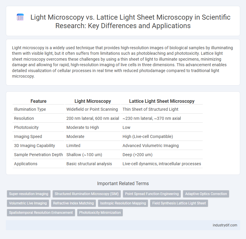
| Feature | Light Microscopy | Lattice Light Sheet Microscopy |
|---|---|---|
| Illumination Type | Widefield or Point Scanning | Thin Sheet of Structured Light |
| Resolution | 200 nm lateral, 600 nm axial | ~230 nm lateral, ~370 nm axial |
| Phototoxicity | Moderate to High | Low |
| Imaging Speed | Moderate | High (Live-cell Compatible) |
| 3D Imaging Capability | Limited | Advanced Volumetric Imaging |
| Sample Penetration Depth | Shallow (~100 um) | Deep (>200 um) |
| Applications | Basic structural analysis | Live-cell dynamics, intracellular processes |
Introduction to Light Microscopy
Light microscopy utilizes visible light and optical lenses to magnify specimens, allowing detailed observation of cellular structures up to approximately 200 nanometers resolution. Traditional light microscopes rely on wide-field illumination, which can cause photobleaching and phototoxicity in live samples, limiting temporal imaging capabilities. Lattice Light Sheet Microscopy (LLSM) improves upon these limitations by using ultrathin sheets of structured light for high-resolution, three-dimensional imaging with reduced photodamage, enabling long-term observation of dynamic biological processes.
Principles of Lattice Light Sheet Microscopy
Lattice Light Sheet Microscopy employs a structured light sheet generated by interfering laser beams to produce an ultrathin illumination plane, reducing phototoxicity and photobleaching in live-cell imaging. Its principle involves selective plane illumination coupled with rapid volumetric imaging, enabling high spatiotemporal resolution of dynamic cellular processes. Unlike conventional light microscopy, this technique minimizes out-of-focus light, enhancing contrast and resolution in three-dimensional samples.
Resolution and Imaging Capabilities
Light microscopy offers a resolution limit of approximately 200 nanometers, constrained by the diffraction of visible light, which restricts its ability to capture fine subcellular structures. Lattice light sheet microscopy, utilizing structured illumination and thin light sheets, achieves superior spatiotemporal resolution with reduced phototoxicity, enabling volumetric imaging of live cells at sub-100 nanometer resolution. This advanced technique provides enhanced imaging capabilities for dynamic processes in three dimensions, surpassing conventional light microscopy in both depth penetration and temporal resolution.
Phototoxicity and Sample Viability
Light microscopy often induces higher phototoxicity due to prolonged exposure and intense illumination, which can compromise sample viability in live-cell imaging. Lattice light sheet microscopy minimizes phototoxic effects by using a thin sheet of light to selectively illuminate the specimen, resulting in reduced photobleaching and enhanced preservation of cellular functions. This technique enables extended time-lapse imaging with improved sample health, critical for observing dynamic biological processes in real time.
Temporal and Spatial Resolution Comparisons
Light microscopy typically offers spatial resolution limited to around 200 nanometers and temporal resolution constrained by frame acquisition speed, often insufficient for capturing rapid cellular dynamics. Lattice Light Sheet Microscopy (LLSM) achieves superior spatial resolution near 100 nanometers and temporal resolution in the range of milliseconds, enabling high-speed, high-contrast imaging of live cells with minimal phototoxicity. LLSM's enhanced temporal and spatial precision allows for detailed visualization of subcellular structures over time, surpassing conventional wide-field and confocal microscopy capabilities.
Applications in Biological Research
Light microscopy remains a foundational tool in biological research for observing cellular structures and processes with high spatial resolution. Lattice light sheet microscopy advances this capability by enabling rapid, high-contrast three-dimensional imaging of live cells and tissues with minimal phototoxicity, making it ideal for dynamic studies at subcellular resolution. Applications such as tracking intracellular trafficking, studying developmental biology, and visualizing protein interactions benefit significantly from the enhanced imaging speed and reduced photodamage offered by lattice light sheet microscopy.
Limitations and Technical Challenges
Light Microscopy faces limitations in spatial resolution and phototoxicity, restricting its application in long-term live-cell imaging. Lattice Light Sheet Microscopy overcomes some of these challenges by providing higher spatial resolution with minimized photodamage, yet it requires complex instrumentation and precise alignment, increasing technical difficulty and cost. Both techniques encounter challenges in balancing imaging speed, resolution, and sample viability, impacting their usability in dynamic biological processes.
Sample Preparation Requirements
Light microscopy typically requires simple sample preparation, such as staining and mounting on glass slides, enabling straightforward visualization of fixed or live specimens. Lattice light sheet microscopy demands more elaborate preparation to maintain sample viability while enabling high-resolution, three-dimensional imaging with minimal phototoxicity, often involving embedding samples in specialized media compatible with rapid light-sheet scanning. The distinctive sample preparation protocols directly influence imaging quality, resolution, and the ability to observe dynamic cellular processes in real-time.
Advancements in Live-Cell Imaging
Lattice light sheet microscopy significantly enhances live-cell imaging by providing higher spatiotemporal resolution while minimizing phototoxicity compared to traditional light microscopy. This technique utilizes ultrathin sheets of light to illuminate specimens, enabling rapid, volumetric imaging of dynamic cellular processes with minimal damage. The resulting improvements facilitate detailed visualization of subcellular structures and real-time tracking of molecular interactions in living cells.
Future Trends in Light Microscopy Technologies
Lattice Light Sheet Microscopy (LLSM) offers unprecedented spatiotemporal resolution and reduced phototoxicity compared to traditional light microscopy, enabling real-time imaging of dynamic cellular processes at subcellular detail. Future trends in light microscopy prioritize integrating adaptive optics with LLSM to correct sample-induced aberrations, enhancing imaging depth and clarity in thick tissues. Advances in computational imaging and machine learning algorithms will further optimize data acquisition and analysis, pushing the boundaries of live-cell imaging and high-throughput biological studies.
Related Important Terms
Super-resolution Imaging
Light Microscopy typically achieves a resolution limit of approximately 200 nanometers due to diffraction constraints, whereas Lattice Light Sheet Microscopy combines structured illumination with selective plane illumination to enable super-resolution imaging with enhanced spatiotemporal resolution and reduced phototoxicity. This advanced technique allows visualization of live cellular processes at subcellular levels, reaching lateral resolutions below 100 nanometers while minimizing photobleaching and providing rapid volumetric imaging.
Structured Illumination Microscopy (SIM)
Structured Illumination Microscopy (SIM) enhances resolution by projecting patterned light onto specimens, surpassing the diffraction limit of conventional light microscopy and enabling detailed visualization of subcellular structures. Lattice Light Sheet Microscopy integrates SIM principles with selective plane illumination, providing rapid, high-contrast 3D imaging with reduced phototoxicity, ideal for live-cell imaging at high spatiotemporal resolution.
Point Spread Function Engineering
Lattice Light Sheet Microscopy achieves superior axial resolution and reduced phototoxicity compared to traditional Light Microscopy by employing engineered point spread functions (PSFs) that create ultrathin, uniform illumination sheets. This PSF engineering facilitates high-speed volumetric imaging with minimal out-of-focus light, enabling precise subcellular structure visualization in living cells.
Adaptive Optics Correction
Adaptive optics correction in light microscopy enhances image resolution by compensating for optical aberrations caused by tissue heterogeneity, but lattice light sheet microscopy (LLSM) integrates adaptive optics more effectively due to its planar illumination and reduced phototoxicity. This integration in LLSM enables high-speed, volumetric imaging with superior spatial accuracy, making it ideal for live-cell and deep-tissue imaging applications.
Volumetric Live Imaging
Light microscopy offers high-resolution imaging but often suffers from phototoxicity and limited volumetric imaging speed in live specimens. Lattice light sheet microscopy overcomes these limitations by providing rapid, high-contrast volumetric live imaging with minimal photodamage, enabling detailed observation of dynamic cellular processes in three dimensions over extended periods.
Refractive Index Matching
Refractive index matching in light microscopy minimizes optical aberrations by aligning the indices of immersion media, coverslips, and specimens to enhance image clarity and resolution. Lattice Light Sheet Microscopy optimizes refractive index matching through its thin, planar illumination, reducing scattering and phototoxicity while enabling high-resolution, 3D imaging of living cells with minimal distortion.
Isotropic Resolution Mapping
Lattice Light Sheet Microscopy offers superior isotropic resolution mapping compared to conventional Light Microscopy by minimizing phototoxicity and photobleaching during high-speed, volumetric imaging of live cells. This advanced technique enables near-uniform axial and lateral resolution, enhancing the visualization of dynamic cellular processes at nanoscale detail.
Field Synthesis Lattice Light Sheet
Light Microscopy offers traditional wide-field imaging with limited axial resolution and phototoxicity risks, while Lattice Light Sheet Microscopy (LLSM) utilizes structured illumination to achieve high spatiotemporal resolution and reduced photodamage. Field Synthesis Lattice Light Sheet enhances LLSM by enabling rapid volumetric imaging with improved light sheet uniformity and minimal out-of-focus excitation, critical for observing dynamic cellular processes in three dimensions.
Spatiotemporal Resolution Enhancement
Lattice Light Sheet Microscopy achieves superior spatiotemporal resolution compared to conventional Light Microscopy by using a thin, structured light sheet that minimizes phototoxicity and photobleaching while enabling rapid volumetric imaging with subcellular detail. This advancement allows for precise observation of dynamic biological processes in living cells at millisecond timescales and submicron spatial resolution, surpassing the diffraction limits inherent in standard widefield or confocal microscopy techniques.
Phototoxicity Minimization
Lattice light sheet microscopy significantly reduces phototoxicity by illuminating specimens with a thin sheet of light, minimizing exposure compared to traditional widefield or confocal light microscopy that generates higher photodamage through intense and prolonged illumination. This method enables long-term imaging of living cells with reduced reactive oxygen species production, preserving cellular physiology and viability.
Light Microscopy vs Lattice Light Sheet Microscopy Infographic