Medical imaging provides detailed visual representations of anatomical structures, essential for diagnosing and monitoring various health conditions. Radiomics enhances this process by extracting quantitative data from medical images, enabling the identification of patterns that may predict disease prognosis or treatment response. Integrating radiomics with traditional imaging techniques improves precision medicine by offering deeper insights into tumor heterogeneity and personalized patient care.
Table of Comparison
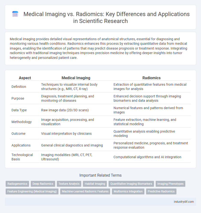
| Aspect | Medical Imaging | Radiomics |
|---|---|---|
| Definition | Techniques to visualize internal body structures (e.g., MRI, CT, X-ray) | Extraction of quantitative features from medical images for analysis |
| Purpose | Diagnosis, treatment planning, and monitoring of diseases | Enhanced decision support through imaging biomarkers and data analysis |
| Data Type | Raw image data (2D/3D scans) | Numerical features and patterns derived from images |
| Methodology | Image acquisition, processing, and visualization | Feature extraction, machine learning, and statistical modeling |
| Outcome | Visual interpretation by clinicians | Quantitative analysis enabling predictive modeling |
| Applications | General clinical diagnostics and imaging | Personalized medicine, prognosis, and treatment response evaluation |
| Technological Basis | Imaging modalities (MRI, CT, PET, Ultrasound) | Computational algorithms and AI integration |
Overview of Medical Imaging and Radiomics
Medical imaging encompasses techniques such as MRI, CT, and ultrasound that visualize internal anatomical structures for diagnostic and therapeutic purposes. Radiomics extracts high-dimensional quantitative features from these medical images, enabling detailed characterization of tissue heterogeneity and disease phenotypes. Integrating radiomics with medical imaging enhances precision medicine by facilitating biomarker discovery and improving clinical decision-making.
Key Technologies in Medical Imaging
Medical imaging employs advanced technologies such as MRI, CT, and ultrasound to capture detailed anatomical and functional information non-invasively. Radiomics enhances these images by extracting quantitative features using machine learning algorithms, enabling the conversion of visual data into high-dimensional, mineable datasets. Key technologies in medical imaging focus on image acquisition, reconstruction, and enhancement, while radiomics emphasizes feature extraction, pattern recognition, and predictive modeling.
Introduction to Radiomics: Definition and Scope
Radiomics is an advanced field within medical imaging that extracts a large number of quantitative features from medical images using data-characterization algorithms. It bridges imaging and personalized medicine by transforming images into high-dimensional data that reveal tumor heterogeneity and underlying pathophysiology. This quantitative analysis enhances diagnostic accuracy, prognosis prediction, and therapeutic decision-making beyond conventional radiological assessments.
Data Acquisition: From Imaging to Radiomics
Medical imaging involves capturing visual representations of the body's interior using modalities such as MRI, CT, and PET, emphasizing high-resolution and standardized data acquisition protocols. Radiomics extends this by extracting quantifiable features from these images, transforming raw imaging data into large-scale, mineable datasets for advanced analysis. Precise image acquisition parameters directly impact the reproducibility and accuracy of radiomic feature extraction, making standardized imaging protocols crucial for effective radiomics application.
Feature Extraction in Radiomics
Feature extraction in radiomics involves quantifying complex patterns from medical images using advanced algorithms, enabling the transformation of pixel data into high-dimensional features that capture tumor heterogeneity. This process surpasses traditional medical imaging analysis by providing objective, reproducible biomarkers that enhance diagnostic precision and prognostic assessment. Techniques such as texture analysis, shape descriptors, and intensity statistics are fundamental in extracting meaningful data to support personalized medicine and predictive modeling.
Applications of Medical Imaging vs Radiomics
Medical imaging techniques such as MRI, CT, and ultrasound provide essential anatomical and functional information for disease diagnosis and treatment monitoring. Radiomics extracts high-dimensional quantitative features from these images, enabling advanced machine learning models to predict disease prognosis, treatment response, and genetic profiles. Applications of radiomics extend precision medicine by offering non-invasive biomarkers for cancer characterization, while traditional medical imaging remains critical for real-time visualization and clinical decision-making.
Role of Artificial Intelligence in Radiomics
Artificial intelligence (AI) in radiomics significantly enhances medical imaging by extracting high-throughput quantitative features beyond visual assessment, enabling precise tumor characterization and personalized treatment strategies. Machine learning algorithms analyze complex imaging data to identify patterns correlated with clinical outcomes, improving diagnostic accuracy and prognostic predictions. AI-driven radiomics transforms traditional imaging into actionable insights, facilitating early disease detection and optimizing patient management.
Benefits and Limitations of Each Approach
Medical imaging provides non-invasive visualization of anatomical structures, offering real-time insights essential for diagnosis and treatment planning but is limited by its qualitative nature and dependency on image quality. Radiomics extracts high-dimensional quantitative features from medical images, enabling personalized medicine through data-driven prognostics and predictive modeling; however, it faces challenges in standardization, reproducibility, and requires large annotated datasets. Combining both approaches can enhance clinical decision-making by integrating visual assessment with robust computational analysis, despite the need for sophisticated computational resources and interdisciplinary expertise.
Integration of Radiomics into Clinical Workflow
Integration of radiomics into clinical workflow enhances medical imaging by extracting high-dimensional data that reveals tumor heterogeneity and microenvironment characteristics beyond conventional imaging. Advanced algorithms and artificial intelligence facilitate the quantification of imaging biomarkers, enabling personalized diagnosis, prognosis, and treatment response prediction. Seamless incorporation of radiomics into existing PACS and electronic health records ensures real-time decision support, promoting precision medicine in oncology and other medical specialties.
Future Trends in Medical Imaging and Radiomics
Future trends in medical imaging and radiomics emphasize integrating artificial intelligence and machine learning to enhance image analysis accuracy and predictive modeling. Advances in multi-modal imaging combine radiomics features from CT, MRI, and PET scans to improve early disease detection and personalized treatment planning. The expansion of big data analytics and deep learning algorithms is driving the development of automated segmentation and classification tools, enabling more precise and scalable diagnostic workflows.
Related Important Terms
Radiogenomics
Radiogenomics integrates radiomic features from medical imaging with genomic data to enhance personalized cancer diagnosis and treatment by identifying molecular characteristics non-invasively. This approach leverages advanced algorithms to correlate imaging phenotypes with genetic profiles, improving predictive accuracy and enabling precision oncology.
Deep Radiomics
Deep Radiomics leverages advanced deep learning algorithms to extract high-dimensional imaging features that surpass traditional Medical Imaging analysis in predicting disease outcomes and treatment responses. This approach integrates convolutional neural networks with radiomic feature extraction, enabling more precise tumor characterization and personalized medicine strategies.
Texture Analysis
Medical imaging techniques provide detailed visual information, while radiomics extracts quantitative features from these images, with texture analysis playing a crucial role in characterizing tissue heterogeneity and tumor microenvironment. Texture analysis metrics such as entropy, uniformity, and gray-level co-occurrence matrices enhance the predictive power of radiomics in oncology diagnostics and treatment response assessment.
Habitat Imaging
Habitat imaging in medical imaging enhances tumor characterization by segmenting heterogeneous regions based on spatial variations in radiomic features, enabling precise mapping of tumor microenvironments. This approach surpasses traditional radiomics by integrating spatial habitat-level data, improving predictive modeling and personalized treatment planning.
Quantitative Imaging Biomarkers
Medical Imaging provides visual representations of anatomical structures using modalities like MRI, CT, and PET, while Radiomics extracts high-dimensional quantitative imaging biomarkers from these images to capture tumor heterogeneity and predict clinical outcomes. Quantitative imaging biomarkers obtained through radiomics enable non-invasive, reproducible assessments that improve diagnostic accuracy, treatment planning, and personalized medicine in oncology.
Imaging Phenotypes
Medical imaging captures detailed anatomical and functional structures, providing high-resolution visualization of tissues, while radiomics extracts quantitative imaging phenotypes through advanced algorithms to identify subtle patterns and biomarkers invisible to the naked eye. Imaging phenotypes derived from radiomics enable more precise disease characterization, prognosis, and treatment response prediction, surpassing conventional imaging interpretation.
Feature Engineering (Medical Imaging)
Medical imaging relies on advanced feature engineering techniques such as texture analysis, shape descriptors, and intensity-based statistics to extract clinically relevant information from visual data. These engineered features serve as the foundation for radiomics, which further quantifies and analyzes high-dimensional imaging biomarkers to improve disease diagnosis and prognosis.
Machine-Learned Radiomic Features
Machine-learned radiomic features extract high-dimensional quantitative data from medical imaging, enabling precise tumor characterization beyond conventional visual assessment. These features integrate complex texture, shape, and intensity patterns, enhancing diagnostic accuracy and predictive modeling in oncology.
Multiomics Integration
Medical imaging provides detailed anatomical and functional data crucial for clinical diagnostics, while radiomics extracts high-dimensional quantitative features from images, enabling deeper phenotypic characterization. Integrating multiomics data--including genomics, proteomics, and metabolomics--with radiomic profiles enhances precision medicine by correlating imaging phenotypes with molecular mechanisms and clinical outcomes.
Predictive Radiomics
Predictive radiomics leverages advanced machine learning algorithms to extract high-dimensional features from medical imaging, enabling precise prognostic assessments beyond traditional imaging interpretation. Integrating radiomic biomarkers with clinical data enhances personalized treatment planning and improves outcome prediction in oncology and other medical fields.
Medical Imaging vs Radiomics Infographic