Conventional microscopy is limited by the diffraction barrier, restricting resolution to approximately 200 nanometers, which hampers the observation of intricate cellular structures. Super-resolution microscopy surpasses this limit by employing advanced techniques such as STED, PALM, and STORM, enabling visualization at the nanoscale with resolutions down to 20 nanometers. This enhanced spatial resolution allows scientists to explore molecular interactions and subcellular components with unprecedented detail, revolutionizing biological and medical research.
Table of Comparison
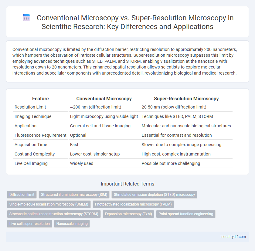
| Feature | Conventional Microscopy | Super-Resolution Microscopy |
|---|---|---|
| Resolution Limit | ~200 nm (diffraction limit) | 20-50 nm (below diffraction limit) |
| Imaging Technique | Light microscopy using visible light | Techniques like STED, PALM, STORM |
| Application | General cell and tissue imaging | Molecular and nanoscale biological structures |
| Fluorescence Requirement | Optional | Essential for contrast and resolution |
| Acquisition Time | Fast | Slower due to complex image processing |
| Cost and Complexity | Lower cost, simpler setup | High cost, complex instrumentation |
| Live Cell Imaging | Widely used | Possible but more challenging |
Introduction to Optical Microscopy Techniques
Optical microscopy techniques encompass conventional microscopy, which relies on visible light and lenses to magnify specimens up to 200 nanometers resolution, and super-resolution microscopy, which surpasses this diffraction limit by employing advanced methods such as STED, PALM, and STORM to achieve resolutions below 20 nanometers. Conventional microscopy is widely used for general biological and material science applications due to its simplicity and real-time imaging capability, whereas super-resolution microscopy enables detailed visualization of molecular structures and interactions at the nanoscale. These advancements in optical microscopy techniques have revolutionized cellular and molecular biology by providing unprecedented spatial resolution and insight into complex biological processes.
Principles of Conventional Microscopy
Conventional microscopy relies on visible light and optical lenses to magnify specimens, with resolution limited by the diffraction limit, typically around 200 nanometers. It uses standard illumination techniques such as bright-field or fluorescence to observe cellular structures but cannot resolve features smaller than half the wavelength of light used. Image formation depends on the interaction of light with the sample, constrained by numerical aperture and wavelength, which restricts its capacity to visualize nanoscale details compared to super-resolution methods.
Limitations of Conventional Microscopy
Conventional microscopy is limited by the diffraction limit of light, which restricts resolution to approximately 200 nanometers, preventing visualization of finer cellular structures. This constraint hampers detailed study of molecular interactions and subcellular components essential in biomedical research. Furthermore, conventional techniques often suffer from low contrast and sensitivity when imaging fluorescently labeled specimens, reducing accuracy in quantitative analysis.
Emergence of Super-Resolution Microscopy
Super-resolution microscopy surpasses the diffraction limit of conventional microscopy, enabling visualization of structures at the nanoscale with unprecedented clarity. Techniques such as STED, PALM, and STORM have revolutionized cellular and molecular imaging by providing resolutions down to 20-30 nanometers. This advancement has accelerated discoveries in fields like neurobiology and virology by revealing intricate details inaccessible through traditional optical microscopy.
Key Super-Resolution Microscopy Techniques
Super-resolution microscopy techniques such as STED (Stimulated Emission Depletion), PALM (Photoactivated Localization Microscopy), and STORM (Stochastic Optical Reconstruction Microscopy) surpass the diffraction limit of conventional light microscopy, achieving resolutions beyond 20 nanometers. These methods utilize precise control of fluorophore activation and emission, enabling detailed visualization of cellular structures at the nanoscale. Compared to traditional wide-field and confocal microscopy, super-resolution approaches provide enhanced spatial resolution critical for molecular and cellular biology research.
Resolution Comparison: Conventional vs Super-Resolution
Conventional microscopy is limited by the diffraction limit of light, typically achieving a maximum resolution of around 200 nanometers, which restricts the visualization of subcellular structures. Super-resolution microscopy techniques, such as STED, PALM, and STORM, break this diffraction barrier, enabling resolutions down to 20-50 nanometers or better. This enhanced resolution allows for detailed imaging of molecular complexes and dynamic processes at the nanoscale, surpassing the capabilities of traditional optical microscopy methods.
Applications in Biological and Material Sciences
Conventional microscopy remains essential for general biological imaging and routine material inspections, providing rapid visualization at diffraction-limited resolution suitable for cell morphology and tissue structure analysis. Super-resolution microscopy techniques, such as STED, PALM, and STORM, enable visualization beyond the diffraction limit, revealing nanoscale details of protein interactions, organelle structures, and nanomaterial properties. These advanced methods have transformed biological research by allowing precise tracking of molecular dynamics and have enhanced material sciences through detailed characterization of nanoscale features in polymers and semiconductor devices.
Sample Preparation and Imaging Requirements
Conventional microscopy typically requires simpler sample preparation, often involving basic staining or fluorescent labeling, suitable for imaging structures above 200 nm resolution. Super-resolution microscopy demands more rigorous sample preparation, including specialized fluorescent probes and precise mounting to minimize photobleaching and maximize signal-to-noise ratio. Imaging with super-resolution techniques also necessitates advanced hardware adaptations, such as high-powered lasers and sensitive detectors, to achieve resolutions down to 20 nm.
Data Analysis and Image Interpretation
Conventional microscopy relies on diffraction-limited resolution, which restricts the ability to accurately analyze and interpret fine structural details at the nanoscale. Super-resolution microscopy surpasses this limit by utilizing techniques such as STED, PALM, and STORM, enabling enhanced spatial resolution and more precise molecular localization. Advanced image processing algorithms and quantitative data analysis in super-resolution microscopy facilitate improved interpretation of complex biological structures and dynamic processes.
Future Perspectives in Microscopy Technologies
Super-resolution microscopy surpasses the diffraction limit of conventional microscopy, enabling visualization of cellular structures at the nanoscale with unprecedented clarity. Emerging techniques such as MINFLUX and expansion microscopy promise further improvements in spatial resolution and imaging speed, facilitating real-time observation of dynamic biological processes. Integrating artificial intelligence and adaptive optics will accelerate data analysis and enhance image quality, paving the way for new discoveries in molecular and cellular biology.
Related Important Terms
Diffraction limit
Conventional microscopy is constrained by the diffraction limit, which restricts spatial resolution to approximately half the wavelength of light used, typically around 200-250 nanometers. Super-resolution microscopy overcomes this barrier by employing techniques such as STED, PALM, and STORM, achieving resolutions down to 20-30 nanometers and enabling visualization of subcellular structures beyond the diffraction limit.
Structured illumination microscopy (SIM)
Conventional microscopy is limited by the diffraction of light, typically achieving a resolution of about 200 nm, whereas super-resolution microscopy techniques like Structured Illumination Microscopy (SIM) surpass this barrier by utilizing patterned illumination and computational reconstruction, boosting resolution to approximately 100 nm. SIM's ability to double spatial resolution while enabling fast image acquisition and multicolor imaging makes it highly effective for live-cell imaging and detailed structural analysis at the nanoscale.
Stimulated emission depletion (STED) microscopy
Stimulated emission depletion (STED) microscopy overcomes the diffraction limit of conventional microscopy by selectively deactivating fluorophores, enabling imaging with nanometer-scale resolution. This super-resolution technique provides detailed visualization of cellular structures and molecular interactions beyond the capabilities of traditional light microscopy.
Single-molecule localization microscopy (SMLM)
Conventional microscopy is limited by diffraction to a resolution of approximately 200 nanometers, whereas super-resolution microscopy techniques like Single-molecule Localization Microscopy (SMLM) achieve nanometer-scale resolution by precisely localizing individual fluorescent molecules. SMLM methods such as PALM and STORM enable visualization of molecular structures and dynamics at the single-molecule level, surpassing the diffraction limit and providing unprecedented insights into cellular processes.
Photoactivated localization microscopy (PALM)
Photoactivated localization microscopy (PALM) surpasses conventional microscopy by achieving nanoscale resolution through the precise activation and localization of photoactivatable fluorescent proteins, enabling visualization of cellular structures at a scale of 10-20 nanometers. This super-resolution technique overcomes the diffraction limit inherent in traditional light microscopy, providing detailed insights into molecular organization and dynamics within biological samples.
Stochastic optical reconstruction microscopy (STORM)
Conventional microscopy is limited by the diffraction limit of light, restricting resolution to approximately 200 nanometers, while super-resolution microscopy techniques like Stochastic Optical Reconstruction Microscopy (STORM) overcome this barrier by localizing individual fluorescent molecules with precision down to 20 nanometers. STORM enables detailed visualization of cellular structures at the nanoscale by repeatedly activating subsets of fluorophores and reconstructing high-resolution images from their stochastic emissions.
Expansion microscopy (ExM)
Expansion microscopy (ExM) physically enlarges biological specimens using a swellable polymer, enabling conventional microscopes to visualize nanoscale structures beyond their diffraction limit. Unlike traditional super-resolution techniques that rely on complex optics and computational reconstruction, ExM achieves high-resolution imaging by isotropic sample expansion, enhancing spatial resolution to approximately 20-70 nanometers with standard fluorescence microscopes.
Point spread function engineering
Point spread function (PSF) engineering in super-resolution microscopy significantly enhances spatial resolution beyond the diffraction limit, enabling visualization of nanoscale biological structures that conventional microscopy cannot resolve. By manipulating the PSF shape and exploiting tailored illumination patterns, super-resolution techniques achieve precise localization of fluorescent molecules, improving imaging accuracy in live-cell and molecular studies.
Live-cell super-resolution
Live-cell super-resolution microscopy surpasses conventional microscopy by breaking the diffraction limit, enabling visualization of cellular structures and dynamic processes at nanometer resolution in real time. Techniques such as STED, PALM, and SIM provide enhanced spatial and temporal resolution while minimizing phototoxicity, facilitating detailed study of molecular interactions in living cells.
Nanoscale imaging
Conventional microscopy is limited by the diffraction limit of light, typically restricting resolution to around 200 nanometers, whereas super-resolution microscopy techniques such as STED, PALM, and STORM enable imaging at the nanoscale, achieving resolutions below 20 nanometers. These advanced methods allow detailed visualization of subcellular structures and molecular interactions that remain unresolved with traditional optical microscopy.
Conventional microscopy vs Super-resolution microscopy Infographic