Microscopy allows detailed visualization of pet specimens' cellular structures using light or electron beams, offering insights into morphology and function. Cryo-electron microscopy enhances this by capturing samples in a near-native frozen state, preserving delicate biological molecules without chemical fixation. This technique provides high-resolution, three-dimensional images critical for studying complex protein assemblies and ultrastructural details in scientific pet research.
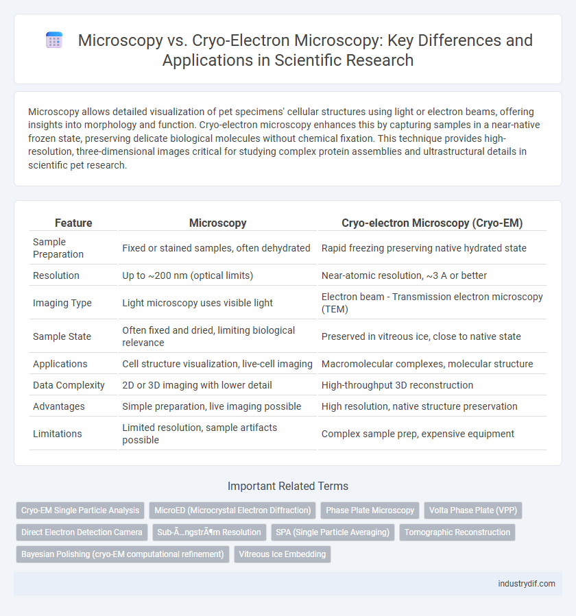
Table of Comparison
| Feature | Microscopy | Cryo-electron Microscopy (Cryo-EM) |
|---|---|---|
| Sample Preparation | Fixed or stained samples, often dehydrated | Rapid freezing preserving native hydrated state |
| Resolution | Up to ~200 nm (optical limits) | Near-atomic resolution, ~3 A or better |
| Imaging Type | Light microscopy uses visible light | Electron beam - Transmission electron microscopy (TEM) |
| Sample State | Often fixed and dried, limiting biological relevance | Preserved in vitreous ice, close to native state |
| Applications | Cell structure visualization, live-cell imaging | Macromolecular complexes, molecular structure |
| Data Complexity | 2D or 3D imaging with lower detail | High-throughput 3D reconstruction |
| Advantages | Simple preparation, live imaging possible | High resolution, native structure preservation |
| Limitations | Limited resolution, sample artifacts possible | Complex sample prep, expensive equipment |
Introduction to Microscopy and Cryo-electron Microscopy
Microscopy encompasses various techniques that utilize light or electron beams to magnify and visualize minute structures, enabling detailed examination of biological and material specimens. Cryo-electron microscopy (cryo-EM) is a specialized form of electron microscopy that involves rapid freezing of samples to preserve their native state without the need for staining or fixation, allowing high-resolution imaging at near-atomic levels. The development of cryo-EM has revolutionized structural biology by providing precise insights into protein complexes, viruses, and cellular organelles that traditional microscopy methods cannot achieve with comparable clarity.
Fundamental Principles of Conventional Microscopy
Conventional microscopy relies on visible light and optical lenses to magnify specimens up to around 1000x resolution, constrained by the diffraction limit of light (~200 nm). It uses transmitted or reflected light to create images of stained or naturally pigmented samples, allowing observation of cellular structures and morphology. However, its fundamental principle is limited in resolving molecular complexes or fine ultrastructural details compared to electron-based methods.
Overview of Cryo-electron Microscopy Techniques
Cryo-electron microscopy (cryo-EM) employs rapid freezing techniques to preserve specimens in a near-native state, enabling high-resolution imaging of biomolecular complexes without the need for crystallization. This method utilizes electron beams to capture detailed structural information, facilitating the study of proteins, viruses, and cellular organelles at atomic or near-atomic resolution. Compared to traditional microscopy, cryo-EM offers superior visualization of dynamic biological processes and complex assemblies under physiological conditions.
Resolution Limits: Microscopy vs Cryo-EM
Conventional light microscopy is limited by the diffraction limit, typically achieving resolutions around 200 nanometers, which restricts detailed visualization of molecular structures. Cryo-electron microscopy (Cryo-EM) surpasses these limits by using electron beams with much shorter wavelengths, enabling near-atomic resolution down to 3-4 angstroms. This enhanced resolution allows Cryo-EM to reveal intricate biomolecular conformations and complexes that are inaccessible to traditional microscopy techniques.
Sample Preparation and Handling Differences
Microscopy techniques differ markedly in sample preparation and handling protocols, with traditional light microscopy often requiring chemical staining and thin sectioning to enhance contrast, while cryo-electron microscopy (cryo-EM) necessitates rapid vitrification of samples to preserve native structures in a near-native hydrated state. Cryo-EM samples are typically flash-frozen on grids under cryogenic conditions to prevent ice crystallization, ensuring high-resolution imaging at molecular scales, unlike conventional microscopy where fixation and dehydration may introduce artifacts. The delicate handling in cryo-EM demands specialized equipment such as plunge freezers and cryo-transfer systems, emphasizing the need for maintaining cryogenic temperatures throughout preparation and imaging workflows.
Imaging Biological Macromolecules: A Comparative Approach
Microscopy techniques such as light microscopy offer lower resolution imaging of biological macromolecules compared to cryo-electron microscopy (cryo-EM), which achieves near-atomic resolution by imaging samples rapidly frozen in vitreous ice. Cryo-EM enables visualization of macromolecular conformations in native-like states without the need for crystallization, facilitating structural studies of dynamic protein complexes and membrane proteins. The ability of cryo-EM to capture heterogeneous states and large assemblies surpasses traditional microscopy, making it a powerful tool for structural biology and drug discovery.
Structural Biology Applications in Research
Microscopy techniques, including light microscopy and electron microscopy, are essential for visualizing cellular structures and macromolecules, while cryo-electron microscopy (cryo-EM) specifically enables high-resolution imaging of biomolecules in near-native states without the need for crystallization. Cryo-EM has revolutionized structural biology by allowing researchers to determine the 3D structures of large protein complexes and membrane proteins at atomic resolution, facilitating drug discovery and understanding protein function. The ability of cryo-EM to capture multiple conformational states expands insights into dynamic biological processes, surpassing traditional microscopy limitations in structural research.
Advantages and Limitations of Each Modality
Light microscopy enables quick visualization of living cells with minimal sample preparation but is limited by a resolution of approximately 200 nanometers due to the diffraction limit of light. Cryo-electron microscopy (Cryo-EM) achieves near-atomic resolution by imaging vitrified samples at cryogenic temperatures, allowing detailed structural analysis of macromolecules in a close-to-native state. However, Cryo-EM requires complex sample preparation, specialized equipment, and is less effective for rapid, live-cell imaging compared to conventional microscopy.
Technological Advances Driving the Field
Microscopy has evolved significantly with the integration of advanced imaging techniques, yet cryo-electron microscopy (cryo-EM) revolutionizes structural biology by enabling near-atomic resolution visualization of biomolecules in their native hydrated states without crystallization. Technological advances such as direct electron detectors, improved image processing algorithms, and phase plate technology have enhanced cryo-EM's resolution and throughput, surpassing limitations of traditional light and electron microscopy. The increasing accessibility of cryo-EM facilities combined with automation and machine learning-driven data analysis accelerates biomolecular research and drug discovery applications.
Future Trends in Microscopy and Cryo-EM
Future trends in microscopy and cryo-electron microscopy (Cryo-EM) emphasize enhanced resolution and real-time imaging capabilities, driven by advances in detector technology and computational algorithms. Integration of artificial intelligence and machine learning accelerates data processing and structural analysis, facilitating more precise molecular insights. Innovations in sample preparation and cryogenic preservation techniques aim to minimize artifacts and expand the applicability of Cryo-EM to dynamic biological processes.
Related Important Terms
Cryo-EM Single Particle Analysis
Cryo-electron microscopy (Cryo-EM) single particle analysis enables high-resolution three-dimensional reconstructions of individual macromolecules by rapidly freezing samples in vitreous ice, preserving their native state without crystallization requirements. This technique surpasses traditional light microscopy in resolving structural details at near-atomic resolution, revolutionizing structural biology and drug design through its ability to visualize dynamic conformations and heterogeneous samples.
MicroED (Microcrystal Electron Diffraction)
MicroED (Microcrystal Electron Diffraction) leverages cryo-electron microscopy to determine atomic structures from nanocrystals that are too small for traditional X-ray crystallography, providing high-resolution data with minimal radiation damage. This technique surpasses conventional microscopy by enabling structural analysis of biomolecules in near-native states, advancing fields like structural biology and drug discovery.
Phase Plate Microscopy
Phase plate microscopy enhances contrast in electron microscopy by manipulating the phase of scattered electrons, enabling detailed visualization of biological specimens without staining. Compared to standard cryo-electron microscopy, phase plate techniques significantly improve image contrast and resolution, facilitating the study of macromolecular complexes in near-native states.
Volta Phase Plate (VPP)
The Volta Phase Plate significantly enhances image contrast in cryo-electron microscopy by inducing a continuous phase shift without the need for defocusing, enabling high-resolution visualization of biological specimens at near-native states. Unlike conventional light microscopy, VPP-equipped cryo-EM overcomes limitations related to sample thickness and electron scattering, providing detailed structural information at the molecular level.
Direct Electron Detection Camera
Direct Electron Detection Cameras in Cryo-electron Microscopy (Cryo-EM) provide superior electron sensitivity and faster frame rates compared to traditional microscopy detectors, enabling enhanced image resolution at near-atomic levels. This technology minimizes electron beam damage while improving signal-to-noise ratio, critical for observing biological macromolecules in their native states.
Sub-Ångström Resolution
Cryo-electron microscopy (cryo-EM) achieves sub-Angstrom resolution by preserving native biomolecular structures in vitrified ice, avoiding artifacts common in traditional microscopy techniques. Advanced direct electron detectors and image processing algorithms enable cryo-EM to resolve atomic details, surpassing the spatial resolution limits of conventional light and electron microscopy.
SPA (Single Particle Averaging)
Microscopy techniques such as Transmission Electron Microscopy (TEM) provide high-resolution structural insights, but Cryo-electron Microscopy (Cryo-EM) coupled with Single Particle Averaging (SPA) significantly enhances molecular imaging by allowing the visualization of biomolecules in near-native states without crystallization. SPA in Cryo-EM improves signal-to-noise ratio through computational alignment and averaging of thousands of particle images, enabling atomic-level resolution of complex protein structures and dynamic conformations.
Tomographic Reconstruction
Tomographic reconstruction in microscopy involves generating three-dimensional images from two-dimensional projections, enabling detailed analysis of complex biological structures. Cryo-electron microscopy (cryo-EM) enhances this process by preserving samples in vitreous ice, reducing radiation damage and improving resolution for precise structural elucidation at near-atomic scales.
Bayesian Polishing (cryo-EM computational refinement)
Bayesian Polishing in Cryo-electron Microscopy enhances particle image alignment and motion correction by applying a probabilistic model that refines individual particle trajectories, significantly improving resolution and structural accuracy compared to traditional Microscopy methods. This computational refinement leverages the Bayesian framework to statistically optimize noise reduction and beam-induced motion artifacts, enabling high-fidelity reconstructions at near-atomic levels.
Vitreous Ice Embedding
Microscopy techniques differ significantly in sample preparation, with traditional electron microscopy often requiring dehydration and heavy-metal staining, whereas cryo-electron microscopy (cryo-EM) preserves biological specimens embedded in vitreous ice, maintaining native hydration and ultrastructure. Vitreous ice embedding in cryo-EM prevents ice crystal formation that can disrupt molecular integrity, enabling high-resolution imaging of macromolecular complexes in near-native states.
Microscopy vs Cryo-electron Microscopy Infographic