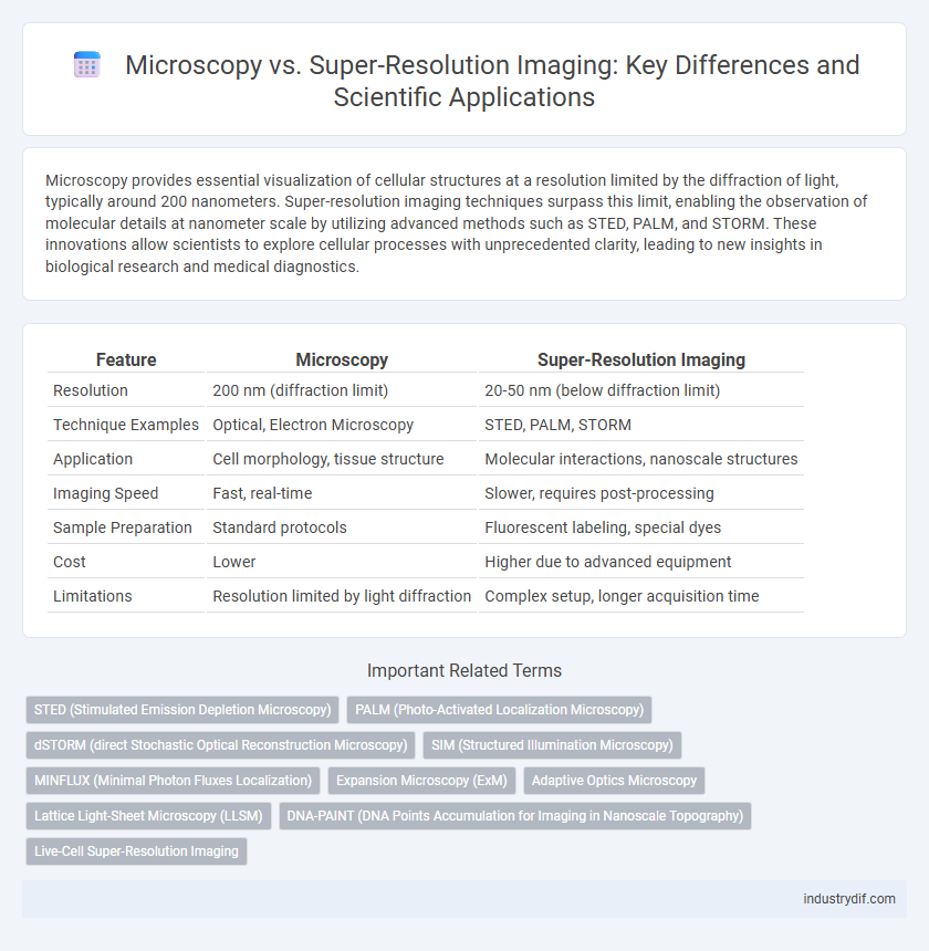
Microscopy provides essential visualization of cellular structures at a resolution limited by the diffraction of light, typically around 200 nanometers. Super-resolution imaging techniques surpass this limit, enabling the observation of molecular details at nanometer scale by utilizing advanced methods such as STED, PALM, and STORM. These innovations allow scientists to explore cellular processes with unprecedented clarity, leading to new insights in biological research and medical diagnostics.
Table of Comparison
| Feature | Microscopy | Super-Resolution Imaging |
|---|---|---|
| Resolution | 200 nm (diffraction limit) | 20-50 nm (below diffraction limit) |
| Technique Examples | Optical, Electron Microscopy | STED, PALM, STORM |
| Application | Cell morphology, tissue structure | Molecular interactions, nanoscale structures |
| Imaging Speed | Fast, real-time | Slower, requires post-processing |
| Sample Preparation | Standard protocols | Fluorescent labeling, special dyes |
| Cost | Lower | Higher due to advanced equipment |
| Limitations | Resolution limited by light diffraction | Complex setup, longer acquisition time |
Introduction to Microscopy and Super-Resolution Imaging
Microscopy enables visualization of structures at the micro- to nanoscale using techniques such as light, electron, and fluorescence microscopy. Super-resolution imaging overcomes the diffraction limit of conventional light microscopy, achieving spatial resolution beyond 200 nanometers by employing methods like STED, PALM, and STORM. These advancements allow detailed analysis of molecular interactions and cellular architecture with unprecedented clarity.
Historical Development of Imaging Techniques
Microscopy has evolved significantly since the 17th century, beginning with Antonie van Leeuwenhoek's single-lens microscopes, which revolutionized biological observation. The diffraction limit posed by light waves constrained resolution until the late 20th century, when super-resolution imaging techniques such as STED, PALM, and STORM emerged, surpassing traditional limits down to tens of nanometers. These advances enabled unprecedented visualization of cellular structures, fueling breakthroughs in cell biology and molecular neuroscience.
Key Principles of Conventional Microscopy
Conventional microscopy relies on the diffraction limit of light, typically around 200 nanometers, which restricts the resolution achievable by standard optical systems. It uses lenses to magnify and resolve specimens based on visible light wavelengths, producing images through transmitted or reflected light. Key principles include the numerical aperture of lenses, illumination intensity, and contrast techniques such as phase contrast or fluorescence to enhance specimen visibility.
Limitations of Traditional Microscopy
Traditional microscopy is constrained by the diffraction limit of light, which caps resolution at approximately 200 nanometers, preventing visualization of subcellular structures with high detail. It often suffers from low contrast and poor depth penetration when imaging thick specimens, limiting its effectiveness in observing dynamic biological processes in vivo. These limitations necessitate advanced techniques like super-resolution imaging that surpass the diffraction barrier, providing nanoscale resolution and enhanced molecular detail.
Emergence and Fundamentals of Super-Resolution Imaging
Super-resolution imaging emerged to overcome the diffraction limit inherent in traditional light microscopy, enabling visualization of structures at the nanoscale with resolution down to tens of nanometers. Techniques such as STED (Stimulated Emission Depletion), PALM (Photoactivated Localization Microscopy), and STORM (Stochastic Optical Reconstruction Microscopy) utilize innovative light manipulation and fluorescent molecule localization strategies to achieve this enhanced resolution. The fundamental principle involves precise spatial control and temporal modulation of fluorescence signals, allowing researchers to reconstruct high-resolution images beyond the capabilities of conventional microscopy.
Comparative Resolution and Imaging Capabilities
Microscopy traditionally offers resolution limited by the diffraction of light to approximately 200 nanometers, whereas super-resolution imaging techniques such as STED, PALM, and STORM achieve resolutions down to 20 nanometers or less by overcoming this diffraction barrier. Super-resolution imaging enables visualization of molecular structures and dynamics at the nanoscale, providing detailed insights unattainable by conventional microscopy. The enhanced resolution and imaging capabilities facilitate advanced studies in cell biology, neuroscience, and materials science, promoting breakthroughs in understanding subcellular processes.
Common Techniques in Super-Resolution Microscopy
Super-resolution microscopy techniques surpass the diffraction limit of traditional light microscopy, enabling visualization of cellular structures at the nanoscale. Common methods include STED (Stimulated Emission Depletion) microscopy, which uses a depletion laser to refine the focal spot size, and PALM (Photo-Activated Localization Microscopy) along with STORM (Stochastic Optical Reconstruction Microscopy), which rely on the precise localization of individual fluorescent molecules. These techniques provide spatial resolutions below 50 nanometers, crucial for detailed studies in cell biology and molecular interactions.
Applications in Biological and Material Sciences
Microscopy techniques enable cellular visualization with spatial resolutions limited by the diffraction barrier, typically around 200 nanometers, essential for routine observation in biology and materials science. Super-resolution imaging surpasses this limit, achieving resolutions down to 20 nanometers or less, facilitating detailed analysis of molecular interactions, protein localization, and nanoscale material structures. Applications in biological sciences include live-cell imaging and tracking protein dynamics, while in material sciences, super-resolution imaging aids in characterizing nanomaterials and defects at an unprecedented scale.
Challenges and Considerations in Advanced Imaging
Microscopy techniques face limitations in spatial resolution due to the diffraction limit of light, restricting the ability to observe nanoscale structures clearly. Super-resolution imaging overcomes these constraints by utilizing methods such as STED, PALM, and STORM, yet they present challenges including photobleaching, extended acquisition times, and complex data analysis. Careful consideration of sample preparation, fluorophore selection, and imaging conditions is essential to optimize image quality and accuracy in advanced imaging applications.
Future Trends in Scientific Imaging Technologies
Emerging advancements in microscopy, such as adaptive optics and AI-driven image reconstruction, are poised to significantly enhance resolution and imaging speed beyond traditional limits. Super-resolution imaging techniques like STED, PALM, and SIM continue to evolve, enabling visualization of molecular processes at nanoscale with increasing accessibility and live-cell compatibility. Future scientific imaging technologies will likely integrate multi-modal approaches, combining super-resolution with real-time data analytics to revolutionize cellular and molecular research.
Related Important Terms
STED (Stimulated Emission Depletion Microscopy)
STED microscopy surpasses conventional microscopy by utilizing a depletion laser to quench fluorophores around the excitation spot, achieving nanometer-scale resolution beyond the diffraction limit. This technique enables detailed visualization of cellular structures and molecular interactions that remain unresolved with traditional fluorescence microscopy.
PALM (Photo-Activated Localization Microscopy)
PALM (Photo-Activated Localization Microscopy) surpasses traditional microscopy by achieving nanoscale resolution through the precise localization of photoactivatable fluorescent proteins, enabling visualization of cellular structures beyond the diffraction limit. This super-resolution imaging technique allows for detailed mapping of molecular distributions with spatial resolutions down to 20 nm, revolutionizing studies in cell biology and molecular neuroscience.
dSTORM (direct Stochastic Optical Reconstruction Microscopy)
dSTORM (direct Stochastic Optical Reconstruction Microscopy) surpasses conventional microscopy by achieving nanometer-scale resolution through the precise localization of individual fluorescent molecules, enabling visualization of cellular structures beyond the diffraction limit. This super-resolution imaging technique relies on the stochastic switching of fluorophores to reconstruct high-resolution images, providing critical insights into molecular organization and dynamics at the nanoscale.
SIM (Structured Illumination Microscopy)
Structured Illumination Microscopy (SIM) enhances spatial resolution by illuminating specimens with patterned light and computationally reconstructing high-resolution images, surpassing the diffraction limit of conventional fluorescence microscopy. SIM achieves lateral resolution improvements to approximately 100 nm, enabling detailed visualization of cellular structures with reduced phototoxicity compared to other super-resolution techniques such as STED or PALM/STORM.
MINFLUX (Minimal Photon Fluxes Localization)
MINFLUX (Minimal Photon Fluxes Localization) achieves nanometer-scale resolution by combining the spatial precision of fluorescence microscopy with minimal photon emission, surpassing traditional microscopy limits. This super-resolution technique enables detailed visualization of molecular structures and dynamics with spatial resolution around 1-3 nm, far beyond conventional diffraction-limited imaging.
Expansion Microscopy (ExM)
Expansion Microscopy (ExM) enhances spatial resolution by physically enlarging biological specimens, enabling nanoscale imaging with conventional microscopes compared to traditional super-resolution techniques like STED or PALM that rely on complex optics and fluorescent labeling. ExM allows detailed visualization of subcellular structures by isotropically expanding samples up to 4-10 times, providing accessible, high-resolution imaging without requiring specialized microscopy setups.
Adaptive Optics Microscopy
Adaptive optics microscopy corrects wavefront distortions in real-time, significantly improving image resolution and contrast beyond conventional microscopy limits. This technique enhances super-resolution imaging by compensating for sample-induced aberrations, enabling detailed visualization of subcellular structures in thick biological tissues.
Lattice Light-Sheet Microscopy (LLSM)
Lattice Light-Sheet Microscopy (LLSM) combines the advantages of traditional microscopy with super-resolution imaging by using thin light sheets created through lattice patterns to minimize phototoxicity and photobleaching while enabling high spatiotemporal resolution. LLSM provides unprecedented 3D visualization of live cellular processes at subcellular scales, surpassing the diffraction limit constraints typical of conventional fluorescence microscopy.
DNA-PAINT (DNA Points Accumulation for Imaging in Nanoscale Topography)
DNA-PAINT (DNA Points Accumulation for Imaging in Nanoscale Topography) surpasses traditional microscopy by achieving nanometer-scale resolution through transient DNA hybridization, enabling detailed visualization of biomolecular structures beyond the diffraction limit. This super-resolution imaging technique combines high specificity and multiplexing capability, making it essential for studying complex cellular architectures and protein interactions at the molecular level.
Live-Cell Super-Resolution Imaging
Live-cell super-resolution imaging utilizes techniques such as STED, PALM, and SIM to surpass the diffraction limit of conventional microscopy, enabling visualization of dynamic cellular processes at nanometer resolution. These methods provide critical insights into protein interactions, organelle dynamics, and intracellular trafficking in living cells without compromising cell viability.
Microscopy vs Super-Resolution Imaging Infographic

 industrydif.com