Traditional microscopy techniques provide valuable insights into cellular structures but are limited by light diffraction, restricting resolution to about 200 nanometers. Super-resolution microscopy overcomes this barrier, allowing visualization of molecular details at the nanoscale with resolutions down to 20 nanometers or less. This enhanced resolution enables more precise study of subcellular components, revolutionizing research in cell biology and neuroscience.
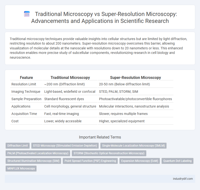
Table of Comparison
| Feature | Traditional Microscopy | Super-Resolution Microscopy |
|---|---|---|
| Resolution Limit | ~200 nm (Diffraction limit) | 20-50 nm (Below diffraction limit) |
| Imaging Technique | Light-based, widefield or confocal | STED, PALM, STORM, SIM |
| Sample Preparation | Standard fluorescent dyes | Photoactivatable/photoconvertible fluorophores |
| Applications | Cell morphology, general structure | Molecular interactions, nanostructure analysis |
| Acquisition Time | Fast, real-time imaging | Slower, requires multiple frames |
| Cost | Lower, widely accessible | Higher, specialized equipment |
Introduction to Microscopy in Scientific Research
Microscopy plays a critical role in scientific research by enabling visualization of structures beyond the resolution of the naked eye. Traditional microscopy techniques, such as light and electron microscopy, provide foundational imaging but are limited by diffraction limits and sample preparation constraints. Super-resolution microscopy overcomes these barriers by employing advanced fluorescent labeling and computational methods, achieving nanometer-scale resolution critical for studying molecular and cellular dynamics.
Fundamentals of Traditional Microscopy Techniques
Traditional microscopy relies on optical lenses to magnify specimens using visible light, limited by the diffraction barrier to a resolution of approximately 200 nanometers. Techniques such as bright-field, phase-contrast, and fluorescence microscopy employ different illumination and contrast methods to enhance specimen visualization without surpassing the diffraction limit. The fundamental constraint in traditional microscopy arises from the wavelength of light, restricting resolution and necessitating alternative approaches for nanoscale imaging.
Limitations of Conventional Microscopy
Conventional microscopy is constrained by the diffraction limit of light, typically around 200 nanometers, restricting the ability to resolve finer cellular structures. This limitation hampers detailed visualization of subcellular components and molecular interactions critical for advanced biological research. Super-resolution microscopy techniques overcome these barriers by enabling imaging at resolutions down to 20 nanometers, facilitating unprecedented insights into molecular organization and dynamics.
Emergence of Super-Resolution Microscopy
Super-resolution microscopy overcomes the diffraction limit of traditional light microscopy, enabling visualization of cellular structures at nanometer resolution. Techniques such as STED, PALM, and STORM have revolutionized biological imaging by providing unprecedented detail at the molecular level. This emergence has significantly advanced the understanding of complex cellular processes that were previously unresolvable with conventional microscopy.
Key Super-Resolution Microscopy Methods
Super-resolution microscopy methods such as STED (Stimulated Emission Depletion), PALM (Photoactivated Localization Microscopy), and STORM (Stochastic Optical Reconstruction Microscopy) surpass the diffraction limit of conventional microscopy, achieving nanoscale resolution below 20 nanometers. These techniques enable precise visualization of subcellular structures by utilizing fluorescent molecules activated or depleted in a controlled manner, enhancing spatial resolution beyond the 200 nm limit of traditional light microscopy. The development of these methods has revolutionized cell biology by allowing dynamic imaging of protein complexes and molecular interactions with unprecedented clarity.
Resolution Comparison: Traditional vs. Super-Resolution
Traditional microscopy techniques, such as optical and electron microscopy, are typically limited by the diffraction limit, restricting resolution to approximately 200 nanometers. Super-resolution microscopy methods, including STED, PALM, and STORM, surpass this barrier by achieving resolutions down to 20-50 nanometers, enabling visualization of subcellular structures previously unresolvable. This enhanced resolution allows for detailed molecular and cellular analysis critical for advanced biomedical research.
Sample Preparation and Imaging Requirements
Traditional microscopy requires relatively simple sample preparation, often involving basic staining and fixation techniques suitable for visualizing general cellular structures. In contrast, super-resolution microscopy demands more sophisticated sample preparation, including fluorescent labeling with specific probes and stringent environmental controls to maintain fluorophore stability and minimize photobleaching. Imaging requirements for super-resolution techniques involve advanced optical setups with precise control over illumination patterns and detector sensitivity to achieve nanometer-scale resolution beyond the diffraction limit of conventional optics.
Applications in Biological and Material Sciences
Traditional microscopy, including optical and electron microscopy, remains essential for observing cellular structures and materials at micrometer to nanometer scales but is limited by diffraction constraints. Super-resolution microscopy techniques, such as STED, PALM, and STORM, enable visualization of subcellular components and nanomaterials with resolutions beyond the diffraction limit, facilitating detailed studies of protein interactions, membrane dynamics, and nanostructured materials. In biological sciences, super-resolution microscopy advances understanding of molecular mechanisms in live cells, while in material sciences, it elucidates nanoscale defects and compositions crucial for developing advanced materials.
Challenges and Future Trends in Microscopy
Traditional microscopy is limited by optical diffraction, restricting resolution to approximately 200 nanometers, which hinders detailed visualization of subcellular structures. Super-resolution microscopy overcomes this barrier, achieving resolution down to 20 nanometers or less, but faces challenges such as photobleaching, complex sample preparation, and high equipment costs. Future trends focus on enhancing temporal resolution, developing label-free super-resolution techniques, and integrating artificial intelligence for automated image analysis and improved data interpretation.
Conclusion: Advancing Scientific Discovery Through Microscopy
Super-resolution microscopy surpasses traditional microscopy by breaking the diffraction limit, enabling visualization of cellular structures at the nanometer scale. This advancement facilitates precise localization of biomolecules and dynamic processes, driving breakthroughs in cell biology, neuroscience, and molecular research. Integrating super-resolution techniques accelerates scientific discovery by providing unprecedented spatial resolution and molecular detail.
Related Important Terms
Diffraction Limit
Traditional microscopy is constrained by the diffraction limit, typically around 200 nanometers, which restricts the resolution of subcellular structures. Super-resolution microscopy techniques, such as STED, PALM, and STORM, surpass this barrier by employing fluorescence localization and patterned illumination, achieving resolutions down to 20 nanometers or less.
STED Microscopy (Stimulated Emission Depletion)
STED microscopy surpasses traditional microscopy by overcoming the diffraction limit, achieving spatial resolutions down to 20-30 nanometers through the selective deactivation of fluorophores using stimulated emission depletion. This technique enables detailed visualization of cellular structures and protein interactions at the nanoscale, providing critical insights unattainable with conventional optical microscopy.
Single-Molecule Localization Microscopy (SMLM)
Traditional microscopy techniques are limited by the diffraction limit of light, typically around 200 nm, which restricts the resolution achievable when studying cellular structures. Single-Molecule Localization Microscopy (SMLM), a super-resolution technique, surpasses this limit by precisely localizing individual fluorescent molecules, achieving nanometer-scale resolution and enabling detailed visualization of molecular arrangements within cells.
PALM (Photoactivated Localization Microscopy)
PALM (Photoactivated Localization Microscopy) surpasses traditional microscopy by achieving nanometer-scale resolution through precise localization of photoactivated fluorophores, enabling visualization of cellular structures beyond the diffraction limit. Unlike conventional light microscopy limited to ~200 nm resolution, PALM provides detailed molecular maps essential for studying complex biological processes at the nanoscale.
STORM (Stochastic Optical Reconstruction Microscopy)
STORM (Stochastic Optical Reconstruction Microscopy) surpasses traditional microscopy by achieving nanoscale resolution down to 20-30 nm, enabling detailed visualization of molecular structures beyond the diffraction limit of light. Unlike conventional optical microscopy limited to approximately 200 nm resolution, STORM employs photo-switchable fluorophores and computational reconstruction to map fluorescent molecules with high spatial precision.
Structured Illumination Microscopy (SIM)
Structured Illumination Microscopy (SIM) surpasses traditional microscopy by doubling resolution to approximately 100 nm, enabling visualization of fine cellular structures beyond the diffraction limit of light. This technique uses patterned illumination and computational reconstruction to enhance spatial resolution while maintaining live-cell imaging compatibility, providing significant advantages over conventional widefield and confocal microscopy.
Point Spread Function (PSF) Engineering
Traditional microscopy is limited by the diffraction of light, resulting in a broader Point Spread Function (PSF) that constrains spatial resolution to approximately 200 nanometers. Super-resolution microscopy employs advanced PSF engineering techniques, such as STED, PALM, and STORM, to modify the PSF shape and size, enabling spatial resolution well below the diffraction limit and allowing visualization of subcellular structures at the nanometer scale.
Expansion Microscopy (ExM)
Expansion Microscopy (ExM) achieves nanoscale resolution by physically enlarging biological specimens through polymer embedding and isotropic expansion, surpassing the diffraction limit constraints of traditional light microscopy. Unlike conventional super-resolution techniques, ExM enables high-resolution imaging using standard optical microscopes, facilitating detailed visualization of subcellular structures with simplified sample preparation and reduced photobleaching.
Quantum Dot Labeling
Quantum dot labeling in super-resolution microscopy provides enhanced photostability and brightness compared to traditional organic dyes used in conventional microscopy, enabling precise molecular localization beyond the diffraction limit. This technique facilitates nanoscale imaging with improved signal-to-noise ratio, crucial for detailed visualization of cellular structures and dynamic processes.
MINFLUX Microscopy
MINFLUX microscopy combines minimal photon fluxes with precise localization techniques, achieving spatial resolutions down to 1-3 nanometers, surpassing traditional diffraction limits inherent in conventional microscopy. This method enables unprecedented visualization of molecular structures and dynamics, revolutionizing studies in cellular biology and nanotechnology compared to classical fluorescence microscopy.
Traditional Microscopy vs Super-Resolution Microscopy Infographic