Microscopes use visible light and lenses to magnify samples, providing detailed images of cells and tissues at relatively lower resolutions. Cryo-electron microscopy (Cryo-EM) utilizes electron beams to visualize specimens frozen at cryogenic temperatures, enabling near-atomic resolution of molecular structures without the need for crystallization. Cryo-EM surpasses traditional microscopy in capturing dynamic biological processes and complex protein assemblies with high precision.
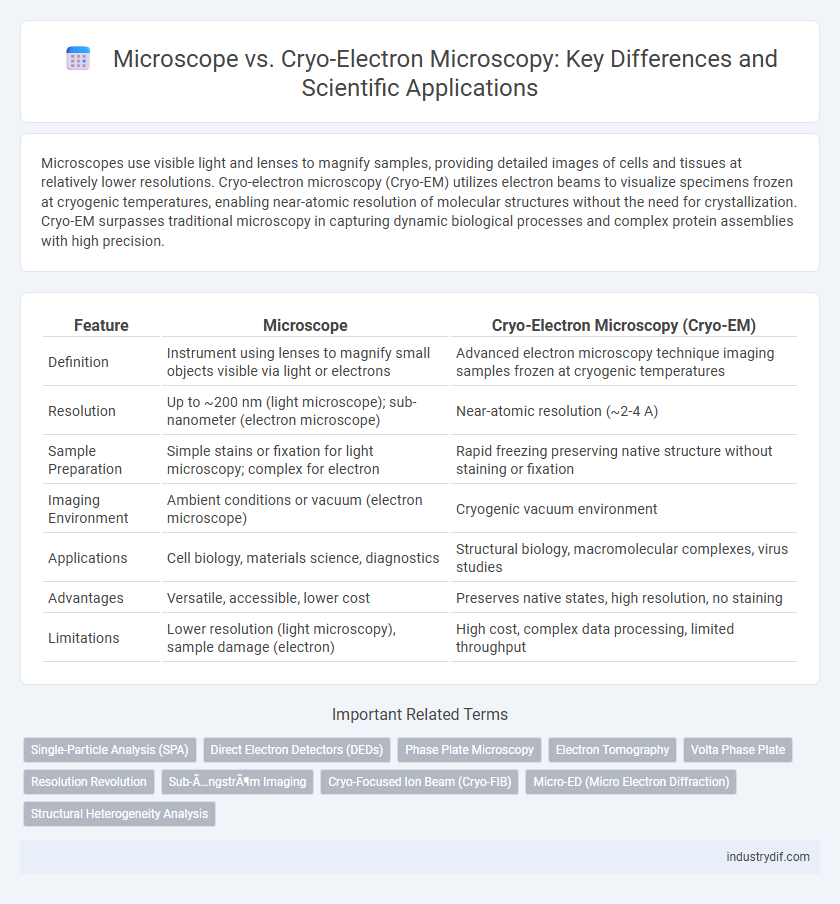
Table of Comparison
| Feature | Microscope | Cryo-Electron Microscopy (Cryo-EM) |
|---|---|---|
| Definition | Instrument using lenses to magnify small objects visible via light or electrons | Advanced electron microscopy technique imaging samples frozen at cryogenic temperatures |
| Resolution | Up to ~200 nm (light microscope); sub-nanometer (electron microscope) | Near-atomic resolution (~2-4 A) |
| Sample Preparation | Simple stains or fixation for light microscopy; complex for electron | Rapid freezing preserving native structure without staining or fixation |
| Imaging Environment | Ambient conditions or vacuum (electron microscope) | Cryogenic vacuum environment |
| Applications | Cell biology, materials science, diagnostics | Structural biology, macromolecular complexes, virus studies |
| Advantages | Versatile, accessible, lower cost | Preserves native states, high resolution, no staining |
| Limitations | Lower resolution (light microscopy), sample damage (electron) | High cost, complex data processing, limited throughput |
Introduction to Microscope Technologies
Microscopes utilize optical lenses to magnify small objects, enabling detailed visualization of specimens primarily at the cellular level. Cryo-electron microscopy (cryo-EM) employs electron beams under cryogenic temperatures, allowing the capture of high-resolution images of biomolecules in their native hydrated states without staining or fixing. The distinct imaging mechanisms of optical microscopes and cryo-EM provide complementary insights essential for advancing molecular and structural biology research.
Fundamentals of Traditional Microscopy
Traditional microscopy relies on visible light and glass lenses to magnify specimens, enabling observation of cellular structures at resolutions typically limited to around 200 nanometers due to diffraction limits. These optical systems use various contrast techniques, such as phase contrast and fluorescence, to enhance image clarity but cannot resolve molecular-level details. In contrast, cryo-electron microscopy (cryo-EM) utilizes electron beams and ultra-low temperatures to visualize biomolecules at near-atomic resolution, bypassing the fundamental resolution limits of light-based microscopes.
Principles of Cryo-Electron Microscopy (Cryo-EM)
Cryo-Electron Microscopy (Cryo-EM) operates by rapidly freezing biological specimens to preserve their native structures without chemical fixation or staining, enabling imaging at near-atomic resolution. The technique employs electron beams transmitted through vitrified samples, minimizing radiation damage and allowing for high-contrast visualization of macromolecular complexes. Advanced image processing algorithms reconstruct three-dimensional structures from two-dimensional projections, providing detailed insights into molecular architecture and dynamics unattainable by conventional light microscopy.
Resolution Comparison: Light Microscopy vs Cryo-EM
Light microscopy typically achieves a resolution limit of around 200 nanometers due to the diffraction limit of visible light, restricting its ability to visualize subcellular structures in fine detail. Cryo-electron microscopy (Cryo-EM) surpasses this barrier, reaching near-atomic resolution at approximately 3-4 angstroms, enabling detailed visualization of biomolecular complexes. This difference in resolution is critical for structural biology, as Cryo-EM provides unprecedented insight into molecular architectures that are inaccessible to traditional light microscopy.
Sample Preparation Techniques
Microscope sample preparation typically involves chemical fixation, dehydration, and staining to enhance contrast, which can alter native biological structures. Cryo-electron microscopy (cryo-EM) employs rapid freezing methods like vitrification, preserving samples in near-native hydrated states without chemical fixatives. This technique minimizes structural distortions, enabling high-resolution imaging of biomolecules and complex assemblies under cryogenic conditions.
Imaging Capabilities and Limitations
Microscopes, including light and electron types, enable visualization of specimens at varying resolutions, with light microscopes limited to about 200 nm resolution due to diffraction limits, while electron microscopes achieve resolutions down to atomic scales. Cryo-electron microscopy (cryo-EM) excels in imaging biomolecules in near-native hydrated states without the need for crystallization, providing high-resolution 3D structures often below 4 A. Limitations of cryo-EM include sample preparation complexity and reduced imaging speed compared to conventional electron microscopy, whereas traditional microscopes may cause sample damage or artifacts due to staining and dehydration.
Applications in Structural Biology
Microscopes enable visualization of cellular components at high resolution, but Cryo-Electron Microscopy (Cryo-EM) offers near-atomic resolution imaging of biomolecules in their native hydrated state, revolutionizing structural biology. Cryo-EM is particularly effective for studying large protein complexes, membrane proteins, and dynamic assemblies that are difficult to crystallize for X-ray crystallography. Its ability to capture multiple conformational states advances understanding of molecular mechanisms and drug design by providing detailed structural insights.
Data Analysis and Interpretation Methods
Microscope data analysis involves traditional image processing techniques such as contrast enhancement, segmentation, and 3D reconstruction, enabling visualization at nanometer resolution. Cryo-electron microscopy (Cryo-EM) requires advanced computational methods including single-particle analysis, maximum-likelihood classification, and cryo-EM map refinement to interpret molecular structures at near-atomic resolution. Machine learning algorithms and automated particle picking have further improved the accuracy and throughput of Cryo-EM data interpretation, distinguishing it from conventional microscopy analysis workflows.
Cost and Accessibility Considerations
Traditional optical microscopes generally have lower upfront costs and are widely accessible for routine laboratory use, making them suitable for educational and clinical environments. Cryo-electron microscopy (cryo-EM) requires significant investment in expensive equipment such as high-end electron microscopes and cryogenic facilities, which limits its accessibility to well-funded research institutions. Despite the high cost, cryo-EM offers unparalleled resolution and molecular detail, justifying its expense in advanced structural biology studies.
Future Trends in Microscopy Technologies
Microscope technologies are rapidly evolving with Cryo-Electron Microscopy (Cryo-EM) emerging as a transformative tool in structural biology, enabling near-atomic resolution imaging of biomolecules in their native states. Future trends emphasize integration of artificial intelligence and machine learning to enhance image reconstruction, data analysis, and resolution capabilities in both traditional microscopes and Cryo-EM systems. Advances in cryo-focused ion beam milling, phase plate technology, and electron detector sensitivity are poised to further revolutionize high-resolution imaging and expand applications across biomedical research and materials science.
Related Important Terms
Single-Particle Analysis (SPA)
Single-particle analysis (SPA) using cryo-electron microscopy (cryo-EM) enables high-resolution 3D reconstructions of macromolecules in near-native states without crystallization, outperforming traditional light microscopes limited by optical diffraction. Cryo-EM SPA captures thousands of particle images rapidly, facilitating detailed structural insights critical for drug design and molecular biology.
Direct Electron Detectors (DEDs)
Direct Electron Detectors (DEDs) significantly enhance Cryo-Electron Microscopy (Cryo-EM) by providing higher frame rates and improved signal-to-noise ratios compared to traditional CCD cameras in conventional microscopes. This advancement enables real-time correction of beam-induced motion and detailed visualization of biological macromolecules at near-atomic resolution.
Phase Plate Microscopy
Phase plate microscopy enhances contrast in electron microscopy by modulating the phase of scattered electrons, enabling visualization of biological specimens with improved detail compared to conventional bright-field techniques. When integrated with cryo-electron microscopy (cryo-EM), phase plates reduce the need for defocusing, thus preserving high-resolution structural information critical for studying macromolecular complexes in their near-native states.
Electron Tomography
Electron Tomography, an advanced technique within Cryo-Electron Microscopy, provides three-dimensional reconstructions of cellular structures at nanometer resolution, surpassing the two-dimensional limitations of traditional light microscopy. Unlike conventional microscopes, Cryo-EM combined with electron tomography enables visualization of molecular assemblies in near-native states, critical for understanding complex biological processes at the ultrastructural level.
Volta Phase Plate
The Volta phase plate enhances contrast in cryo-electron microscopy (cryo-EM) by modulating the phase shift of electrons, enabling high-resolution imaging of biological macromolecules without the need for staining. Unlike traditional optical microscopes, cryo-EM with the Volta phase plate allows for visualization of specimens in their native state at near-atomic resolution, revolutionizing structural biology.
Resolution Revolution
Cryo-electron microscopy (cryo-EM) has revolutionized structural biology by achieving near-atomic resolution, surpassing the limitations of traditional light microscopes constrained by diffraction limits. The Resolution Revolution in cryo-EM enables visualization of biomolecules in native states with resolutions often better than 3 A, facilitating detailed understanding of molecular mechanisms.
Sub-Ångström Imaging
Microscope techniques such as cryo-electron microscopy (cryo-EM) achieve sub-Angstrom imaging resolution by directly imaging biomolecules in vitrified ice without staining, enabling atomic-level structural analysis. Unlike traditional light or electron microscopes limited by diffraction and sample preparation artifacts, cryo-EM leverages advanced detectors and image processing to visualize molecular conformations near 1 A resolution.
Cryo-Focused Ion Beam (Cryo-FIB)
Cryo-Focused Ion Beam (Cryo-FIB) technology enables precise milling of frozen biological samples, facilitating high-resolution imaging in Cryo-Electron Microscopy (Cryo-EM) by preserving native structures without chemical fixation. This advanced sample preparation technique overcomes limitations of traditional microscopy by producing thin lamellae essential for detailed visualization of cellular and molecular complexes at near-atomic resolution.
Micro-ED (Micro Electron Diffraction)
Micro-ED (Micro Electron Diffraction) combines the high-resolution capabilities of cryo-electron microscopy with electron diffraction to analyze nano-sized crystalline samples at atomic resolution, overcoming limitations of traditional light microscopes. This technique enables structural determination of biomolecules from micro- and nano-crystals that are too small for X-ray crystallography, providing critical insights in structural biology and materials science.
Structural Heterogeneity Analysis
Microscope techniques, including light microscopy, provide limited resolution for structural heterogeneity analysis, whereas Cryo-Electron Microscopy (Cryo-EM) offers near-atomic resolution enabling detailed visualization of molecular conformations and dynamic states. Cryo-EM's ability to capture multiple structural states from heterogeneous samples allows for precise identification and quantification of conformational diversity in macromolecular complexes.
Microscope vs Cryo-Electron Microscopy Infographic