X-ray crystallography provides high-resolution structural data by analyzing crystalized molecules, enabling detailed atomic models but requiring well-ordered crystals. Cryo-electron microscopy allows visualization of biological macromolecules in near-native states without crystallization, excelling in studying large complexes and dynamic conformations. Both techniques contribute complementary insights, with X-ray crystallography offering precision in atomic positioning and cryo-EM enabling analysis of flexible or heterogeneous samples.
Table of Comparison
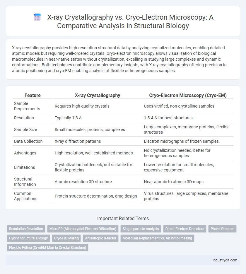
| Feature | X-ray Crystallography | Cryo-Electron Microscopy (Cryo-EM) |
|---|---|---|
| Sample Requirements | Requires high-quality crystals | Uses vitrified, non-crystalline samples |
| Resolution | Typically 1-3 A | 1.5-4 A for best structures |
| Sample Size | Small molecules, proteins, complexes | Large complexes, membrane proteins, flexible structures |
| Data Collection | X-ray diffraction patterns | Electron micrographs of frozen samples |
| Advantages | High resolution, well-established methods | No crystallization needed, better for heterogeneous samples |
| Limitations | Crystallization bottleneck, not suitable for flexible proteins | Lower resolution for small molecules, expensive equipment |
| Structural Information | Atomic resolution 3D structure | Near-atomic to atomic 3D maps |
| Common Applications | Protein structure determination, drug design | Virus structures, large complexes, membrane proteins |
Introduction to X-ray Crystallography and Cryo-EM
X-ray Crystallography involves diffracting X-rays through crystallized molecules to determine atomic structures with high resolution, commonly used for proteins and small molecules. Cryo-Electron Microscopy (Cryo-EM) captures images of rapidly frozen specimens at near-native states, enabling visualization of large biomolecular complexes without crystallization. Both techniques provide complementary insights into molecular architecture, with X-ray Crystallography excelling in atomic detail and Cryo-EM offering structural data on heterogeneous or flexible assemblies.
Principles and Mechanisms of X-ray Crystallography
X-ray crystallography utilizes the diffraction of X-rays through a crystalline lattice to generate electron density maps, revealing atomic structures with high resolution. The method relies on Bragg's law to interpret diffraction patterns produced by constructive interference of X-rays scattered by the periodic arrangement of atoms. Precise phase determination and Fourier transformation are essential for reconstructing three-dimensional molecular structures from the diffraction intensities.
Fundamentals of Cryo-Electron Microscopy
Cryo-Electron Microscopy (Cryo-EM) fundamentally relies on the rapid freezing of biological samples to preserve their native hydrated state, allowing visualization without the need for crystallization required in X-ray Crystallography. By capturing multiple two-dimensional images at different angles, Cryo-EM reconstructs high-resolution three-dimensional structures, enabling detailed analysis of macromolecular complexes, viruses, and membrane proteins. Its capability to study large biomolecules in near-native conditions provides key insights into functional conformations unattainable with traditional crystallographic methods.
Sample Preparation Requirements: Crystals vs Vitrification
X-ray crystallography requires the formation of high-quality crystalline samples, which can be challenging and time-consuming, limiting its applicability to molecules that readily form ordered crystals. Cryo-electron microscopy (cryo-EM) relies on vitrification, where samples are rapidly frozen in a thin layer of amorphous ice, preserving native structures without the need for crystallization. The vitrification process in cryo-EM enables the analysis of heterogeneous and large macromolecular complexes that are often unsuitable for crystallographic approaches.
Resolution Comparison: X-ray Crystallography vs Cryo-EM
X-ray crystallography typically achieves atomic resolutions around 1-2 A, providing highly detailed structural information of crystallized molecules. Cryo-electron microscopy (Cryo-EM) has advanced significantly, now routinely reaching near-atomic resolution of 2-4 A for large macromolecular complexes without the need for crystallization. While X-ray crystallography excels in resolving small molecules and well-ordered crystals, Cryo-EM offers superior capabilities for imaging heterogeneous or flexible specimens at moderate to high resolution.
Structural Data Output and Interpretation
X-ray crystallography provides high-resolution atomic models by analyzing diffraction patterns from crystalline samples, enabling detailed visualization of molecular structures. Cryo-electron microscopy (cryo-EM) generates three-dimensional reconstructions from vitrified specimens, capturing multiple conformational states and larger macromolecular complexes without the need for crystallization. Structural data from X-ray crystallography often yields precise atomic coordinates, while cryo-EM facilitates interpretation of dynamic assemblies and flexible regions, offering complementary insights into biomolecular architecture.
Advantages and Limitations of Each Technique
X-ray crystallography provides high-resolution atomic structures of crystallizable biomolecules, enabling detailed analysis of molecular interactions and conformations, but its limitation lies in the requirement for well-ordered crystals, which can be challenging for large or flexible proteins. Cryo-electron microscopy (Cryo-EM) excels in imaging macromolecular complexes and membrane proteins without crystallization, allowing structural determination of heterogeneous and dynamic samples, though it generally offers lower resolution compared to X-ray crystallography and requires extensive computational processing. Both techniques complement each other by balancing resolution, sample state, and complexity, critical for advancing structural biology and drug discovery.
Applications in Structural Biology and Drug Discovery
X-ray crystallography provides high-resolution atomic structures crucial for understanding protein-ligand interactions in drug discovery, enabling precise drug design targeting specific molecular sites. Cryo-electron microscopy excels in visualizing large macromolecular complexes and membrane proteins in near-native states without crystallization, expanding structural biology's capacity to study dynamic conformations and transient states. Together, these technologies offer complementary insights that accelerate the identification and optimization of therapeutic compounds.
Technological Advances and Future Trends
X-ray crystallography remains a cornerstone in high-resolution structural determination with improvements in detector sensitivity and synchrotron sources enhancing data accuracy. Cryo-electron microscopy (cryo-EM) has witnessed rapid technological advances, including direct electron detectors and advanced image processing algorithms, enabling near-atomic resolution without the need for crystallization. Future trends highlight integrated hybrid approaches combining crystallography and cryo-EM data to resolve complex biomolecular structures with increased speed and precision.
Choosing the Right Method for Your Research
X-ray crystallography provides atomic-resolution structures ideal for studying well-ordered crystalline samples, making it optimal for analyzing protein-ligand interactions and enzyme mechanisms. Cryo-electron microscopy (Cryo-EM) excels in visualizing large, heterogeneous complexes and membrane proteins without requiring crystallization, enabling structural determination of dynamic and flexible biomolecules. Selecting the appropriate method depends on sample type, resolution needs, and the biological question, with crystallography favored for high-resolution detail and Cryo-EM preferred for complex or non-crystallizable specimens.
Related Important Terms
Resolution Revolution
X-ray crystallography achieves atomic resolution by analyzing diffraction patterns from crystalline samples, whereas cryo-electron microscopy (Cryo-EM) has undergone a resolution revolution, enabling near-atomic resolution imaging of non-crystalline biomolecules in native-like states. Advances in direct electron detectors and image processing algorithms have made Cryo-EM a powerful tool for resolving complex macromolecular structures that are challenging for crystallography.
MicroED (Microcrystal Electron Diffraction)
MicroED (Microcrystal Electron Diffraction) surpasses traditional X-ray crystallography by enabling structural analysis of nanocrystals that are too small for X-ray diffraction, utilizing electron beams to provide higher resolution data with minimal radiation damage. Cryo-Electron Microscopy enhances this approach by preserving samples in vitreous ice, maintaining native-like conformations and allowing detailed visualization of biomolecular structures at near-atomic resolution.
Single-particle Analysis
X-ray crystallography provides atomic-resolution structures by analyzing crystallized biomolecules, while cryo-electron microscopy (cryo-EM) excels in single-particle analysis, capturing heterogeneous, non-crystalline specimens in near-native states without the need for crystallization. Cryo-EM's advancements in direct electron detectors and image processing algorithms enable high-resolution reconstructions of macromolecular complexes, overcoming limitations posed by crystal packing and allowing structural insights into dynamic and flexible biomolecules.
Direct Electron Detectors
Direct electron detectors significantly enhance Cryo-Electron Microscopy by providing improved signal-to-noise ratios and faster frame rates, enabling high-resolution imaging of biological macromolecules without crystallization. Unlike X-ray crystallography, which relies on crystalline samples and indirect detection, the integration of direct electron detectors in Cryo-EM allows for the visualization of flexible and heterogeneous structures in near-native states.
Phase Problem
X-ray crystallography faces the intrinsic phase problem due to the loss of phase information during diffraction, necessitating methods like multiple isomorphous replacement or molecular replacement to reconstruct electron density maps. Cryo-electron microscopy bypasses this challenge by directly imaging particles in ice at near-native states, enabling phase information retrieval without reliance on crystallization or phase estimation techniques.
Hybrid Structural Biology
X-ray crystallography provides atomic resolution structures by analyzing diffraction patterns of crystallized molecules, while cryo-electron microscopy offers 3D reconstructions of biomolecules in near-native states without crystallization. Hybrid structural biology integrates these techniques to leverage crystallography's high resolution and cryo-EM's ability to visualize large, flexible complexes, enhancing the accuracy and completeness of macromolecular models.
Cryo-FIB Milling
Cryo-FIB milling enables precise thinning of vitrified biological samples for Cryo-Electron Microscopy, overcoming thickness limitations inherent to X-ray Crystallography which requires crystalline samples. This technique facilitates high-resolution structural analysis of native macromolecular complexes in near-physiological states without crystallization artifacts.
Anisotropic B-factor
Anisotropic B-factors in X-ray crystallography provide detailed directional atomic displacement information crucial for understanding molecular dynamics at high resolution, while cryo-electron microscopy typically yields isotropic B-factors due to lower resolution and averaging effects. The enhanced directional sensitivity of anisotropic B-factors improves structural refinement accuracy, facilitating precise modeling of protein flexibility and conformational changes.
Molecular Replacement vs. Ab initio Phasing
Molecular replacement in X-ray crystallography relies on known homologous structures as models to solve phase problems, enabling rapid structure determination when similar templates exist. Cryo-electron microscopy's ab initio phasing does not require prior structural models, using direct image reconstruction from raw particle projections to determine novel molecular architectures at near-atomic resolution.
Flexible Fitting (CryoEM-Map to Crystal Structure)
Flexible fitting in Cryo-Electron Microscopy (CryoEM) involves refining atomic models obtained from X-ray crystallography to better conform to CryoEM density maps, improving structural accuracy in regions with conformational heterogeneity. This technique leverages molecular dynamics simulations and real-space refinement algorithms to reconcile discrepancies between high-resolution crystal structures and lower-resolution CryoEM maps, enabling detailed visualization of dynamic protein complexes.
X-ray Crystallography vs Cryo-Electron Microscopy Infographic