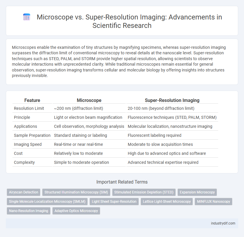
Microscopes enable the examination of tiny structures by magnifying specimens, whereas super-resolution imaging surpasses the diffraction limit of conventional microscopy to reveal details at the nanoscale level. Super-resolution techniques such as STED, PALM, and STORM provide higher spatial resolution, allowing scientists to observe molecular interactions with unprecedented clarity. While traditional microscopes remain essential for general observation, super-resolution imaging transforms cellular and molecular biology by offering insights into structures previously invisible.
Table of Comparison
| Feature | Microscope | Super-Resolution Imaging |
|---|---|---|
| Resolution Limit | ~200 nm (diffraction limit) | 20-100 nm (beyond diffraction limit) |
| Principle | Light or electron beam magnification | Fluorescence techniques (STED, PALM, STORM) |
| Applications | Cell observation, morphology analysis | Molecular localization, nanostructure imaging |
| Sample Preparation | Standard staining or labeling | Fluorescent labeling required |
| Imaging Speed | Real-time or near real-time | Moderate to slow acquisition times |
| Cost | Relatively low to moderate | High due to advanced optics and software |
| Complexity | Simple to moderate operation | Advanced technical expertise required |
Defining Traditional Microscopy
Traditional microscopy involves the use of optical instruments that employ lenses to magnify small objects, typically providing resolution limits around 200 nanometers due to the diffraction limit of visible light. Conventional microscopes rely on light wavelength and numerical aperture to determine their resolving power, often restricting the observation of subcellular structures. This foundational technique serves as a contrast point for super-resolution imaging methods that surpass diffraction barriers to reveal finer molecular details.
Principles of Super-Resolution Imaging
Super-resolution imaging overcomes the diffraction limit of conventional microscopes by utilizing techniques such as stimulated emission depletion (STED), photoactivated localization microscopy (PALM), and structured illumination microscopy (SIM) to achieve nanometer-scale resolution. These methods exploit fluorescence properties and precise spatial or temporal modulation of light to reconstruct images with enhanced detail beyond standard optical limits. The integration of advanced algorithms with optical manipulation enables visualization of cellular structures and molecular interactions at unprecedented resolution.
Resolution Limits: Microscopes vs. Super-Resolution
Conventional optical microscopes are limited by the diffraction limit, typically around 200 nanometers, restricting their ability to resolve structures smaller than this threshold. Super-resolution imaging techniques, such as STED, PALM, and STORM, surpass this boundary by exploiting fluorescence and computational methods to achieve resolutions down to 20-30 nanometers. These advancements enable detailed visualization of subcellular components, providing insights unattainable with traditional microscopy.
Key Technologies in Super-Resolution Imaging
Super-resolution imaging surpasses the diffraction limit of conventional optical microscopes by utilizing techniques such as stimulated emission depletion (STED), photoactivated localization microscopy (PALM), and stochastic optical reconstruction microscopy (STORM). These technologies employ precise control of fluorescent molecules and advanced computational algorithms to achieve nanoscale resolution, enabling visualization of cellular structures beyond 20 nanometers. Integration of adaptive optics and real-time image reconstruction further enhances image clarity and temporal resolution in super-resolution imaging systems.
Sample Preparation: Conventional vs. Advanced Techniques
Sample preparation for conventional microscopy typically involves fixation, staining, and embedding processes that preserve cellular structures but may limit resolution. Advanced super-resolution imaging techniques require more precise and often gentler sample preparation methods, such as the use of fluorescent probes and specialized mounting media, to enhance image clarity and minimize photobleaching. These refined protocols enable the capture of nanometer-scale details essential for high-resolution visualization of molecular interactions.
Imaging Speed and Throughput Comparison
Super-resolution imaging techniques such as STED, PALM, and STORM offer significantly higher spatial resolution than conventional microscopy but often at the cost of slower imaging speed due to complex image acquisition and reconstruction processes. In contrast, traditional fluorescence and confocal microscopes provide faster imaging throughput suitable for live-cell applications but lack the nanoscale resolution achieved by super-resolution methods. Balancing imaging speed and throughput with resolution remains critical in selecting the optimal microscopy approach for specific scientific investigations.
Applications in Life Sciences and Materials Research
Microscope and super-resolution imaging techniques revolutionize visualization in life sciences by enabling detailed study of cellular structures beyond the diffraction limit, crucial for understanding molecular interactions and disease mechanisms. Super-resolution methods such as STED, PALM, and STORM provide nanoscale resolution, facilitating advanced materials research through precise characterization of nanomaterials, polymers, and semiconductor devices. These imaging technologies enhance biomolecular imaging and structural analysis, driving innovations in drug development and nanotechnology.
Quantitative Analysis Capabilities
Super-resolution imaging surpasses traditional microscopy by providing nanoscale spatial resolution, essential for precise quantitative analysis of molecular interactions and cellular structures. Techniques such as STED, PALM, and STORM enable accurate measurement of protein distribution and dynamics, which are limited by the diffraction barrier in conventional microscopy. Quantitative data derived from super-resolution imaging facilitates deeper insights into subcellular processes, enhancing the reliability of biological and biochemical research.
Artifacts and Limitations of Each Approach
Microscopes often produce artifacts such as optical aberrations and diffraction limits, restricting resolution to approximately 200 nanometers, while super-resolution imaging techniques, including STED and PALM, overcome these barriers but introduce artifacts like photobleaching and reconstruction errors. Limitations of conventional microscopy include limited depth penetration and lower contrast, whereas super-resolution methods require complex sample preparation and extensive computational processing, potentially causing inaccuracies. Both approaches must balance artifact management and technique-specific constraints to ensure precise nanoscale imaging.
Future Trends in Scientific Imaging
Super-resolution imaging techniques like STED and PALM are transforming scientific research by surpassing the diffraction limit of conventional microscopes, enabling visualization of cellular structures at the nanometer scale. Future trends emphasize integrating AI-driven image analysis with advanced optics to enhance resolution, reduce imaging time, and increase quantification accuracy. Innovations in adaptive optics and label-free super-resolution methods promise to expand accessibility and applicability in live-cell and in vivo imaging studies.
Related Important Terms
Airyscan Detection
Airyscan detection in super-resolution imaging enhances spatial resolution beyond the diffraction limit of conventional microscopes by utilizing a detector array that captures spatial information from the Airy disk pattern. This technology improves signal-to-noise ratio and enables detailed visualization of subcellular structures with up to 1.7x higher resolution compared to traditional confocal microscopy.
Structured Illumination Microscopy (SIM)
Structured Illumination Microscopy (SIM) enhances conventional microscopy by using patterned light to surpass the diffraction limit, achieving approximately twice the resolution of standard light microscopy, around 100 nm. This super-resolution technique enables detailed visualization of subcellular structures with faster acquisition times and lower phototoxicity compared to other super-resolution methods like STED or PALM.
Stimulated Emission Depletion (STED)
Stimulated Emission Depletion (STED) microscopy surpasses traditional optical microscopes by breaking the diffraction limit, enabling imaging at nanometer resolution. By selectively deactivating fluorophores around the focal spot, STED achieves super-resolution that reveals molecular structures with unprecedented clarity.
Expansion Microscopy
Expansion microscopy enables nanoscale visualization by physically enlarging biological samples, overcoming the diffraction limit inherent in traditional light microscopes and facilitating detailed imaging of molecular structures. This method, combined with super-resolution imaging techniques, provides enhanced spatial resolution critical for studying complex cellular processes at a molecular level.
Single Molecule Localization Microscopy (SMLM)
Single Molecule Localization Microscopy (SMLM) enhances traditional microscope capabilities by surpassing the diffraction limit to achieve nanometer-scale resolution through precise localization of fluorescent molecules. This super-resolution imaging technique provides unprecedented insights into molecular structures and dynamics, revolutionizing fields such as cell biology and neuroscience.
Light Sheet Super-Resolution
Light Sheet Super-Resolution Imaging enhances spatial resolution beyond the diffraction limit by selectively illuminating thin sample sections, reducing phototoxicity and photobleaching compared to conventional confocal microscopes. This technique enables high-speed, volumetric imaging of live cells and tissues with subcellular detail, ideal for dynamic biological processes analysis.
Lattice Light-Sheet Microscopy
Lattice Light-Sheet Microscopy (LLSM) offers superior spatiotemporal resolution compared to conventional microscopes by utilizing structured light sheets to minimize phototoxicity and photobleaching, enabling live-cell imaging at subcellular levels. This technique surpasses traditional super-resolution imaging methods in preserving cellular physiology while delivering dynamic three-dimensional visualization of molecular processes with nanoscale precision.
MINFLUX Nanoscopy
MINFLUX nanoscopy surpasses conventional super-resolution techniques by combining minimal photon fluxes with precise localization, achieving nanometer-scale resolution beyond the diffraction limit of traditional microscopes. This method enables visualization of molecular structures and dynamics with unprecedented spatial and temporal resolution, revolutionizing cellular and molecular biology research.
Nano-Resolution Imaging
Nano-resolution imaging techniques, such as stimulated emission depletion (STED) microscopy and structured illumination microscopy (SIM), surpass traditional optical microscopes by breaking the diffraction limit to achieve resolutions below 100 nanometers. These super-resolution methods enable detailed visualization of molecular structures and dynamic cellular processes at the nanoscale, revolutionizing fields like molecular biology and nanotechnology.
Adaptive Optics Microscopy
Adaptive optics microscopy enhances super-resolution imaging by correcting optical aberrations in real-time, enabling visualization of cellular structures at nanoscale resolution with improved clarity and depth. This technology surpasses conventional microscopes by compensating for sample-induced distortions, thereby achieving higher spatial resolution and precise imaging in complex biological tissues.
Microscope vs Super-Resolution Imaging Infographic