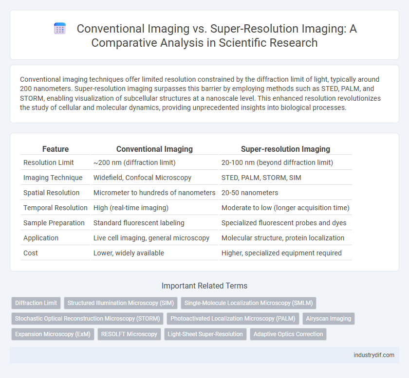
Conventional imaging techniques offer limited resolution constrained by the diffraction limit of light, typically around 200 nanometers. Super-resolution imaging surpasses this barrier by employing methods such as STED, PALM, and STORM, enabling visualization of subcellular structures at a nanoscale level. This enhanced resolution revolutionizes the study of cellular and molecular dynamics, providing unprecedented insights into biological processes.
Table of Comparison
| Feature | Conventional Imaging | Super-resolution Imaging |
|---|---|---|
| Resolution Limit | ~200 nm (diffraction limit) | 20-100 nm (beyond diffraction limit) |
| Imaging Technique | Widefield, Confocal Microscopy | STED, PALM, STORM, SIM |
| Spatial Resolution | Micrometer to hundreds of nanometers | 20-50 nanometers |
| Temporal Resolution | High (real-time imaging) | Moderate to low (longer acquisition time) |
| Sample Preparation | Standard fluorescent labeling | Specialized fluorescent probes and dyes |
| Application | Live cell imaging, general microscopy | Molecular structure, protein localization |
| Cost | Lower, widely available | Higher, specialized equipment required |
Introduction to Imaging Modalities in Science
Conventional imaging techniques, such as confocal and wide-field microscopy, offer spatial resolutions limited by the diffraction limit of light, typically around 200 nanometers. Super-resolution imaging methods, including STED, PALM, and STORM, surpass this barrier by employing precise localization of fluorescent molecules or patterned illumination, achieving resolutions down to 20 nanometers or less. These advancements enable detailed visualization of cellular structures and molecular interactions, fundamentally enhancing scientific research in biology, material science, and nanotechnology.
Principles of Conventional Imaging
Conventional imaging relies on the diffraction limit determined by the wavelength of light and the numerical aperture of the microscope lens, restricting resolution to roughly 200 nanometers. This principle is based on capturing light waves emitted or reflected by a sample and forming an image through lenses without overcoming light scattering or diffraction constraints. As a result, fine structural details below the diffraction limit remain unresolved in conventional optical microscopy.
Fundamentals of Super-resolution Imaging
Super-resolution imaging surpasses the diffraction limit of conventional optical microscopy by utilizing techniques such as stimulated emission depletion (STED), photoactivated localization microscopy (PALM), and stochastic optical reconstruction microscopy (STORM). These methods rely on precise control of fluorescent molecules to achieve nanometer-scale spatial resolution, enabling visualization of cellular structures and molecular interactions with unprecedented detail. By exploiting photo-switchable fluorophores and nonlinear optical effects, super-resolution imaging transforms fundamental light-matter interactions to break traditional resolution barriers.
Key Technologies in Conventional Imaging
Conventional imaging relies on technologies such as optical microscopy, electron microscopy, and X-ray imaging, which primarily capture images limited by the diffraction limit of light, typically around 200 nanometers. Key components include lenses, detectors, and illumination systems designed to maximize resolution within physical constraints. These methodologies form the foundation for observing biological samples, materials, and structures at micro to nanoscale but are inherently restricted by resolution barriers that super-resolution techniques aim to overcome.
Breakthrough Techniques in Super-resolution Imaging
Breakthrough techniques in super-resolution imaging, such as STED (Stimulated Emission Depletion) microscopy, PALM (Photoactivated Localization Microscopy), and STORM (Stochastic Optical Reconstruction Microscopy), surpass conventional imaging limits by resolving structures at the nanoscale below the diffraction limit of light. These advanced methods utilize precise control of fluorescent probes and novel computational algorithms to reconstruct high-resolution images with molecular-level detail. Super-resolution imaging enables unprecedented visualization of cellular components, facilitating significant advancements in molecular biology, neuroscience, and medical diagnostics.
Resolution Limits: Conventional vs Super-resolution
Conventional imaging techniques, constrained by the diffraction limit, typically achieve spatial resolutions around 200-300 nanometers, restricting the ability to visualize sub-cellular structures. Super-resolution imaging surpasses this limit by employing methods such as STED, PALM, and STORM, achieving resolutions down to 10-20 nanometers. This enhanced resolution enables detailed observation of molecular interactions and nanoscale biological processes previously inaccessible with conventional microscopy.
Applications of Conventional Imaging in Research
Conventional imaging techniques, such as brightfield microscopy and confocal microscopy, remain essential tools in cell biology, histology, and pathology for visualizing tissue architecture and cellular morphology. These methods enable researchers to analyze large sample areas rapidly with sufficient resolution for studying cell populations, tissue organization, and standard fluorescent labeling. Despite resolution limits around 200 nanometers, conventional imaging supports quantitative analyses of protein localization, cell counting, and structural abnormalities critical for developmental biology and disease modeling.
Advancements Enabled by Super-resolution Imaging
Super-resolution imaging overcomes the diffraction limit of conventional microscopy, enabling visualization of cellular structures at nanometer-scale resolution. Techniques such as STED, PALM, and STORM provide unprecedented detail in molecular localization, facilitating breakthroughs in understanding protein interactions and subcellular dynamics. These advancements have transformed fields like neurobiology and cell biology, revealing intricate biological processes previously obscured.
Data Analysis and Interpretation Challenges
Conventional imaging techniques often face limitations in resolution and signal-to-noise ratio, leading to challenges in accurately analyzing densely packed molecular structures and dynamic cellular processes. Super-resolution imaging overcomes diffraction limits but introduces complex artifacts and requires advanced computational algorithms for precise localization and quantification of fluorescent signals. Data interpretation in super-resolution demands robust statistical methods to differentiate true biological events from noise, increasing the necessity for specialized software and expertise in image reconstruction and validation.
Future Trends in Scientific Imaging Technologies
Future trends in scientific imaging technologies emphasize the integration of artificial intelligence and machine learning to enhance both conventional and super-resolution imaging techniques. Advances in adaptive optics and novel fluorescent probes are driving improvements in spatial resolution and temporal dynamics, enabling real-time visualization at the nanoscale. Emerging multimodal imaging platforms combine data from super-resolution microscopy with electron microscopy and spectroscopy, providing unprecedented insights into complex biological structures and materials.
Related Important Terms
Diffraction Limit
Conventional imaging techniques are constrained by the diffraction limit, which restricts spatial resolution to approximately half the wavelength of light used, typically around 200-300 nanometers. Super-resolution imaging methods, such as STED, PALM, and STORM, overcome this barrier by exploiting fluorescent molecule behavior and computational algorithms to achieve resolutions below 20 nanometers, enabling visualization of cellular structures at the molecular level.
Structured Illumination Microscopy (SIM)
Conventional imaging techniques are limited by the diffraction limit of light, resulting in resolutions typically around 200 nm, whereas Structured Illumination Microscopy (SIM) achieves super-resolution by projecting patterned illumination and computationally reconstructing images to approximately double the resolution to 100 nm. SIM enables detailed visualization of subcellular structures and dynamic processes in live cells with less phototoxicity compared to other super-resolution methods like STED or PALM/STORM.
Single-Molecule Localization Microscopy (SMLM)
Single-Molecule Localization Microscopy (SMLM) surpasses conventional imaging by achieving nanoscale resolution through precise localization of individual fluorescent molecules, enabling visualization of cellular structures beyond the diffraction limit. Unlike traditional imaging techniques limited to ~200 nm resolution, SMLM attains resolution down to 10-20 nm, significantly enhancing molecular and structural analysis in biological research.
Stochastic Optical Reconstruction Microscopy (STORM)
Conventional imaging techniques are limited by diffraction, restricting resolution to approximately 200 nanometers, whereas Stochastic Optical Reconstruction Microscopy (STORM) surpasses this limit by localizing individual fluorescent molecules with nanometer precision. STORM enables visualization of cellular structures with resolutions down to 20 nanometers, facilitating unprecedented insights into molecular organization and dynamics.
Photoactivated Localization Microscopy (PALM)
Photoactivated Localization Microscopy (PALM) surpasses conventional imaging techniques by achieving nanometer-scale resolution through the precise localization of photoactivatable fluorescent proteins, enabling visualization of cellular structures beyond the diffraction limit. This super-resolution imaging approach provides unprecedented spatial detail critical for studying molecular interactions and dynamic processes in live cells.
Airyscan Imaging
Airyscan imaging enhances resolution beyond conventional confocal microscopy by utilizing a 32-channel GaAsP detector array, which enables improved signal-to-noise ratio and spatial resolution up to 140 nm. This super-resolution technique allows for detailed visualization of subcellular structures with higher sensitivity and faster acquisition times compared to standard imaging methods.
Expansion Microscopy (ExM)
Conventional imaging techniques are limited by the diffraction limit of light, typically resolving structures no smaller than 200 nm, whereas Expansion Microscopy (ExM) physically enlarges biological samples by embedding them in a swellable polymer, enabling nanoscale resolution with standard microscopes. ExM enhances visualization of subcellular structures such as synaptic proteins and cytoskeletal elements, achieving effective resolutions down to 20-70 nm without requiring specialized super-resolution equipment.
RESOLFT Microscopy
RESOLFT microscopy surpasses conventional imaging by breaking the diffraction limit through reversible photoswitchable fluorescent proteins, enabling nanoscale resolution in live-cell imaging. This technique achieves spatial resolution down to 20-30 nanometers, significantly improving the visualization of molecular structures compared to traditional fluorescence microscopy limited to ~200 nanometers.
Light-Sheet Super-Resolution
Light-sheet super-resolution imaging enhances spatial resolution beyond the diffraction limit by selectively illuminating thin specimen planes, reducing phototoxicity and photobleaching typical in conventional imaging. This technique enables high-contrast, volumetric visualization of live cells with subcellular detail, outperforming traditional wide-field and confocal microscopy in speed and sensitivity.
Adaptive Optics Correction
Adaptive optics correction significantly enhances super-resolution imaging by compensating for optical aberrations in real-time, thereby improving resolution beyond the diffraction limit compared to conventional imaging techniques. This technology enables clearer visualization of fine cellular structures, advancing biological and medical research through more precise data acquisition.
Conventional Imaging vs Super-resolution Imaging Infographic